. 2012 Oct 15;84(8):1007-13.
doi: 10.1016/j.bcp.2012.07.014. Epub 2012 Jul 25.

Copper influx transporter 1 is required for FGF, PDGF and EGF-induced MAPK signaling

Affiliations

Copper influx transporter 1 is required for FGF, PDGF and EGF-induced MAPK signaling

Cheng-Yu Tsai et al. Biochem Pharmacol. .

Abstract

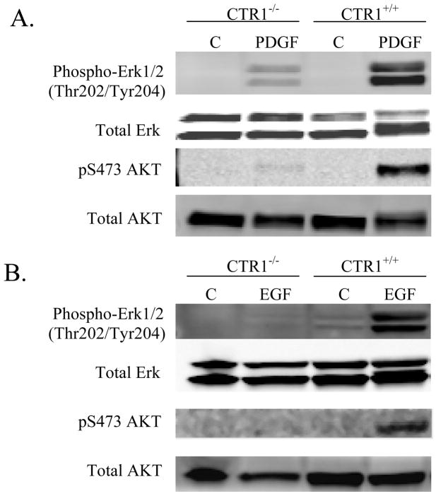
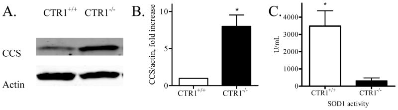
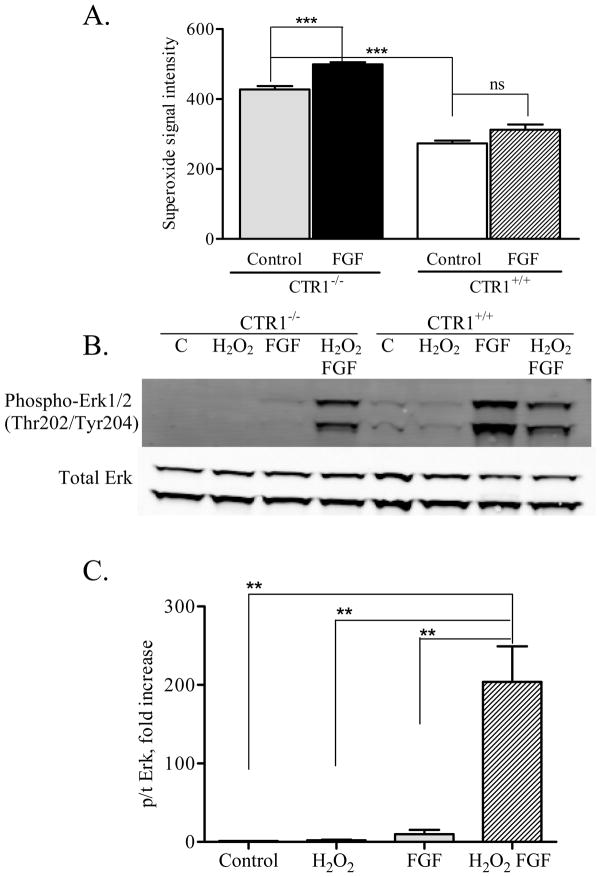
Copper transporter 1 (CTR1) is the major copper (Cu) influx transporter in mammalian cells. We report here that CTR1 is required for the activation of signaling to the MAPK pathway by the ligands of three major receptor tyrosine kinases (RTK) including FGF, PDGF and EGF. Induction of Erk1/2 phosphorylation was compared in isogenic wild type CTR1(+/+) and CTR1(-/-) cells. Whereas all three ligands increased pErk1/2 in the CTR1(+/+) cells, they failed to do this in CTR1(-/-) cells. While FGF did not enhance the phosphorylation of AKT in the CTR1(+/+) cells, both PDGF and EGF increased pAKT in the CTR1(+/+) but not CTR1(-/-) cells. The deficit in Erk1/2 phosphorylation in the CTR1(-/-) cells was rescued by adding Cu to the medium, and it was induced in CTR1(+/+) cells by treatment with a Cu chelator. Intracellular Cu availability was reduced in the CTR1(-/-) cells as reflected by increased expression of the Cu chaperone CCS. The failure of RTK-induced signaling to both Erk1/2 and AKT suggested the presence of a Cu-dependent step upstream of Ras. The Cu-dependent enzyme SOD1 is responsible for generating the hydrogen peroxide in response to RTK activation that serves to inhibit phosphatases that normally limit RTK signaling. SOD1 activity was reduced by a factor of 17-fold in the CTR1(-/-) cells, and addition of hydrogen peroxide restored signaling. We conclude that Cu acquired from CTR1 is required for signaling in pathways regulated by RTKs that play major roles in development and cancer.


Figures

Figure 1
Figure 1
Erk1/2 phosphorylation in response to FGF stimulation. A. CTR1−/− and CTR1+/+ cells were treated with FGF 1 ng/mL for the indicated time points. The immunoblot shown is representative of 3 independent experiments (C, untreated control; S, cells serum starved overnight). B. Quantification of pERK1/2 protein levels in CTR1−/− cells. C. Quantification of pErk1/2 levels in CTR1+/+ cells. Untreated control (white bar), serum starvation (black bar), FGF 1 ng/mL 5 min (gray bar), FGF 1 ng/mL 15 min (hatched bar). Data from immunoblots were normalized to total Erk1/2and expressed as fold increase over untreated control samples (n=3); bars, SE; *, p<0.05, between FGF treatment and untreated control. D. Representative western blot showing failure of FGF to enhance Mek1/2 phosphorylation in the CTR1−/− cells. E. Quantification of FGF-induced phosphorylation of Mek1/2 in CTR1+/+ versus CTR1−/− cells from a total of 4 western blot analyses. Mean untreated control in CTR1−/− (white bar); FGF 1 ng/mL 15 min in CTR1−/− (black bar); untreated control in CTR1+/+ (gray bar); FGF 1 ng/mL 15 min in CTR1+/+ (hatched bar). Vertical, bars, ± SEM; *, p<0.05,
Figure 2
Figure 2
Erk1/2 and AKT phosphorylation in response to PDGF and EGF. A. CTR1−/− and CTR1+/+ cells were treated with PDGF 50 ng/mL for 15 min. B. CTR1−/− and CTR1+/+ cells were treated with EGF 5 ng/mL for 15 min. Immunoblots shown are representative of 3 independent experiments.
Figure 3
Figure 3
Cu deficiency and SOD dysfunction in the CTR1−/− cells A. Western blot documenting an increase in CCS expression in the CTR1−/− cells. The immunoblot shown is representative of 3 independent experiments. B. Quantification of CCS protein level normalized to actin in the CTR1+/+ cells and CTR1−/− cells (n=3); bars, ± SEM.*p<0.05. C. SOD1 activity in the CTR1+/+ cells and CTR1−/− cells (n=3); bars, ± SEM.*p<0.05.
Figure 4
Figure 4
Signaling to Erk1/2 is regulated by the intracellular Cu level. A. FGF-induced phosphorylation of Erk1/2 was restored when CTR1−/− were treated with 200 μM CuSO4 18 h prior to a 15 min exposure to FGF 1 ng/mL. Untreated control (white bar), Cu (black bar), FGF 1 ng/mL 15 min (gray bar), Cu supplement prior to FGF stimulation (hatched bar). B. Exposure to Cu chelator BCS inhibits FGF-induced phosphorylation of Erk1/2 in CTR1+/+ cells. Untreated control (white bar), BCS (black bar), FGF 1 ng/mL 15 min (gray bar), Cu depletion prior to FGF stimulation (hatched bar). For each panel the immunoblots shown are representative of 3 independent experiments. Vertical bars, ± SEM.
Figure 5
Figure 5
Cu deficiency impedes superoxide metabolism. A. Endogenous and stimulated level of superoxide in the paired CTR1 cells. Untreated CTR1 +/+ cells (white bar), FGF treated CTR1 −/− cells (hatched bar), untreated CTR1−/− cells (gray bar), FGF treated CTR1 +/+ cells (black bar), (n=6). B. H2O2 restores FGF-induced phosphorylation of Erk1/2 in CTR1−/− cells. The immunoblot shown is representative of 3 independent experiments. C. Quantification of pERK1/2 levels in CTR1−/− cells. Mean untreated control (white bar); H2O2 (black bar); FGF 1 ng/mL 15 min (gray bar); 100 μmol/L H2O2 30 min prior to FGF stimulation (hatched bar). Bars are mean ± SEM. **p<0.01; ***p<0.001.
Figure 6
Figure 6
Proposed Model. Binding of a growth factor to its RTK activates Rac which then causes NADPH oxidases to generate superoxide. CTR1 is required to provide Cu to SOD1 so that it can dismute superoxide into H2O2. H2O2 oxidizes and inhibits PTPases that normally counteract the phosphorylation cascade triggered by the RTK.

References

    1. Rhee SG. Cell signaling. H2O2, a necessary evil for cell signaling. Science. 2006;312:1882–3. - PubMed
    1. Tonks NK. Redox redux: revisiting PTPs and the control of cell signaling. Cell. 2005;121:667–70. - PubMed
    1. Rhee SG, Kang SW, Jeong W, Chang TS, Yang KS, Woo HA. Intracellular messenger function of hydrogen peroxide and its regulation by peroxiredoxins. Curr Opin Cell Biol. 2005;17:183–9. - PubMed
    1. Ushio-Fukai M. Redox signaling in angiogenesis: role of NADPH oxidase. Cardiovascular research. 2006;71:226–35. - PubMed
    1. Rae TD, Schmidt PJ, Pufahl RA, Culotta VC, O’Halloran TV. Undetectable intracellular free copper: the requirement of a copper chaperone for superoxide dismutase. Science. 1999;284:805–8. - PubMed

Publication types

MeSH terms