Comparative Study
. 2013;42(4):31627230.
doi: 10.1259/dmfr/31627230. Epub 2012 Jul 27.

MRI characteristics of rheumatoid arthritis in the temporomandibular joint

Affiliations
Comparative Study

MRI characteristics of rheumatoid arthritis in the temporomandibular joint

K Kretapirom et al. Dentomaxillofac Radiol. 2013.

Abstract

Objectives: The aim of this study was to investigate characteristic MRI findings of rheumatoid arthritis (RA) in the temporomandibular joints (TMJs).

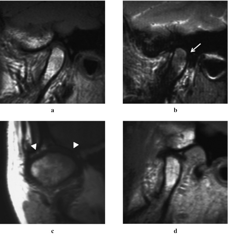
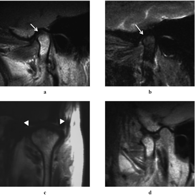
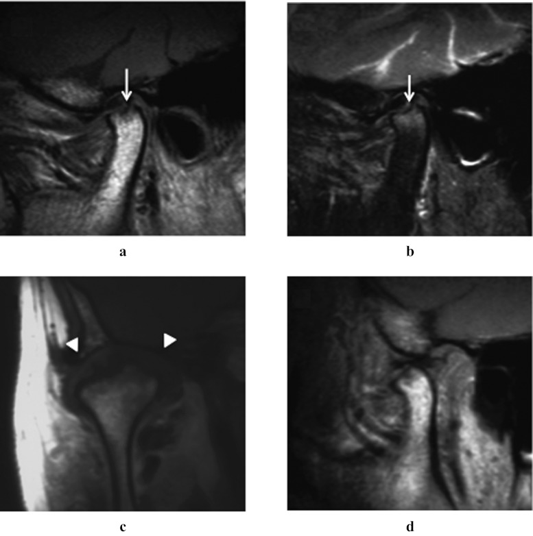
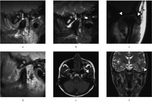
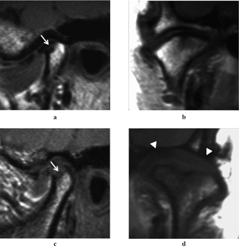
Methods: 61 patients (122 TMJs) with RA in the TMJ and 50 patients (100 TMJs) with temporomandibular disorder (TMD) were included in this study. MR images of these patients were assessed by two oral radiologists for the presence or absence of osseous changes, disc displacement, joint effusion and synovial proliferation. These findings were compared between the two patient groups.

Results: Osseous changes in the condyle and articular eminence/fossa in the RA patient group were significantly more frequent than in the TMD patient group, and were often very severe. Joint effusion was also significantly more frequent in the RA patient group. Synovial proliferation was found in all TMJs in the RA patient group, whereas it was very uncommon in the TMD patient group.

Conclusions: Severe osseous changes in the condyle and synovial proliferation were considered characteristic MRI findings of RA in the TMJs.


Figures

Figure 1
Figure 1
Schematic drawing showing four types of osseous changes in the condyle. Type I, a condyle showing abnormal signal intensity of the bone marrow without erosion or absorption. Type II, a condyle with erosion in the cortex. Type III, a condyle with bone absorption extending within half of the condyle. Type IV, a condyle with bone absorption extending over half of the condyle
Figure 2
Figure 2
A 45-year-old female with rheumatoid arthritis. Sagittal proton density-weighted image (closed-mouth position). (a) Sagittal T2 weighted image with fat saturation (closed-mouth position). (b) Coronal proton density-weighted image (closed-mouth position). (c) Sagittal proton density-weighted image (open-mouth position). T2 weighted MR image of the right temporomandibular joint revealed inhomogeneous high-signal intensity in the condyle (b, arrow, Type I). Synovial proliferation was also revealed (c, arrowheads)
Figure 3
Figure 3
A 68-year-old female with rheumatoid arthritis. (a) Sagittal proton density-weighted image (closed-mouth position). (b) Sagittal T2 weighted image with fat saturation (closed-mouth position). (c) Coronal proton density-weighted image (closed-mouth position). (d) Sagittal proton density-weighted image (open-mouth position). MR images of the left temporomandibular joint revealed erosion in the cortex of the condyle (a,b, arrows, Type II) and synovial proliferation (c, arrowheads)
Figure 4
Figure 4
A 50-year-old female with rheumatoid arthritis. (a) Sagittal proton density-weighted image (closed-mouth position). (b) Sagittal T2 weighted image with fat saturation (closed-mouth position). (c) Coronal proton density-weighted image (closed- mouth position). (d) Sagittal proton density-weighted image (open-mouth position). MR images of the right temporomandibular joint revealed bone absorption extending within half of the condyle (a,b, arrows, Type III) and remarked synovial proliferation (c, arrowheads)
Figure 5
Figure 5
A 32-year-old female with rheumatoid arthritis. (a) Sagittal proton density-weighted image (closed-mouth position). (b) Sagittal T2 weighted image with fat saturation (closed-mouth position). (c) Coronal proton density-weighted image (closed-mouth position). (d) Sagittal proton density-weighted image (open-mouth position). (e) Axial T1 -weighted image. (f) Post-contrast coronal T1 weighted image. MR images of the left temporomandibular joint revealed bone absorption extending over half of the condyle (a,b, arrows, Type IV) and remarked synovial proliferation (c,e,f, arrowheads). Joint effusion was also found. On the post-contrast image (f), the proliferated synovium of the left TMJ showed strong enhancement
Figure 6
Figure 6
Comparison of MRI findings between rheumatoid arthritis (RA) and temporomandibular disorder (TMD). (a,b) A 47-year-old female with TMD. (c,d) A 43-year-old female with RA. (a,c) Sagittal proton density-weighted image (closed-mouth position). (b, d) Coronal proton density-weighted image (closed-mouth position). MR images of the temporomandibular joint revealed bone absorption extending within half of the condyle in both patients (a,c, arrows, Type III). However, synovial proliferation was evident in the RA patient only (d, arrowheads)

References

    1. Vliet Vlieland TP, Buitenhuis NA, van Zeben D, Vandenbroucke JP, Breedveld FC, Hazes JM. Sociodemographic factors and the outcome of rheumatoid arthritis in young women. Ann Rheum Dis 1994; 53: 803–806 - PMC - PubMed
    1. Fleishaker DL, Garcia Meijide JA, Petrov A, Kohen MD, Wang X, Menon S, et al. Maraviroc, a chemokine receptor-5 antagonist, fails to demonstrate efficacy in the treatment of patients with rheumatoid arthritis in a randomized, double-blind placebo-controlled trial. Arthritis Res Ther 2012; 14: R11. - PMC - PubMed
    1. Gylys-Morin VM, Graham TB, Blebea JS, Dardzinski BJ, Laor T, Johnson ND, et al. Knee in early juvenile rheumatoid arthritis: MR imaging findings. Radiology 2001; 220: 696–706 - PubMed
    1. Sugimoto H, Takeda A, Hyodoh K. Early-stage rheumatoid arthritis: prospective study of the effectiveness of MR imaging for diagnosis. Radiology 2000; 216: 569–757 - PubMed
    1. Marbach JJ, Spiera H. Rheumatoid arthritis of the temporomandibular joints. Ann Rheum Dis 1967; 26: 538–543 - PMC - PubMed

Publication types