. 2012 Sep 20;489(7416):400-5.
doi: 10.1038/nature11375. Epub 2012 Jul 29.

Structural plasticity and dynamic selectivity of acid-sensing ion channel-spider toxin complexes

Affiliations

Structural plasticity and dynamic selectivity of acid-sensing ion channel-spider toxin complexes

Isabelle Baconguis et al. Nature. .

Abstract

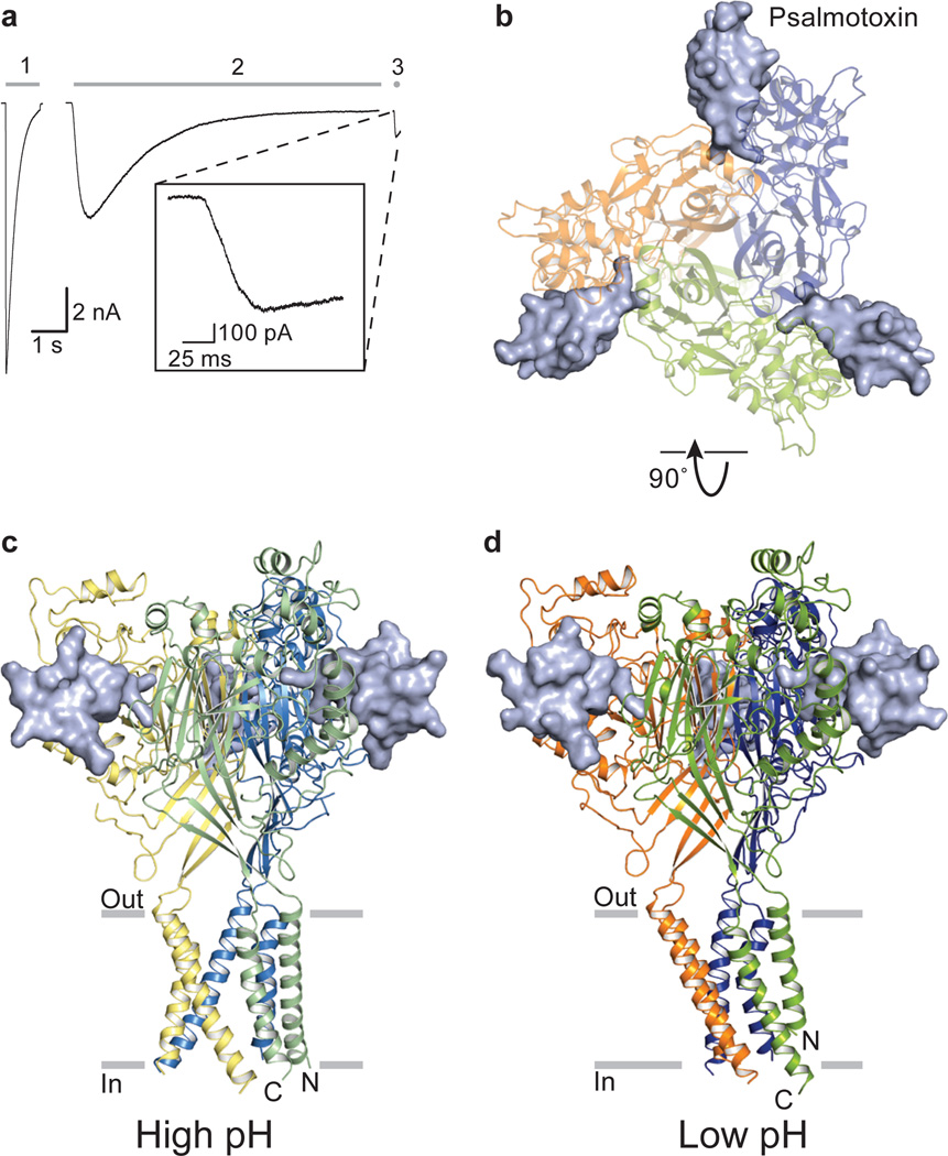
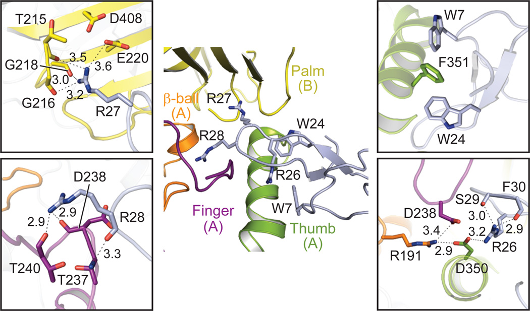
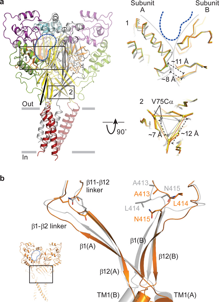
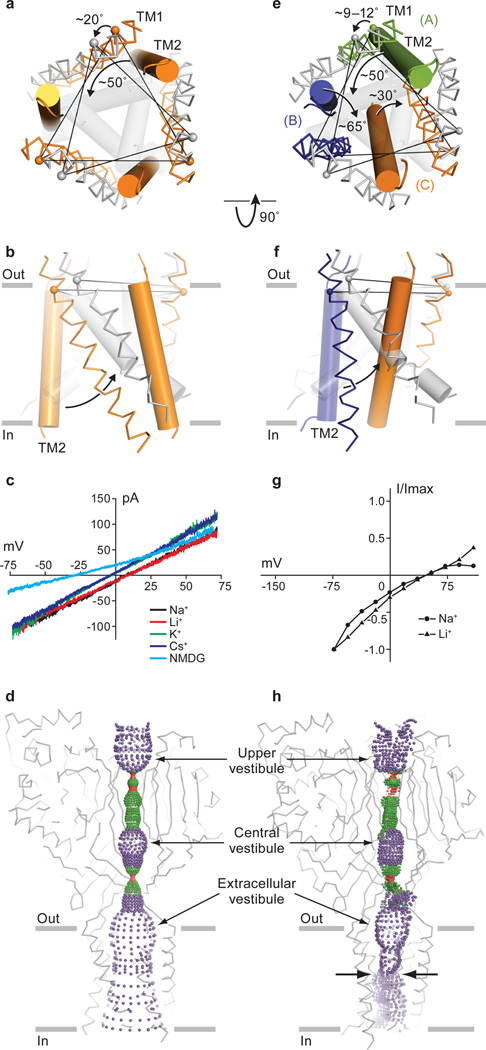
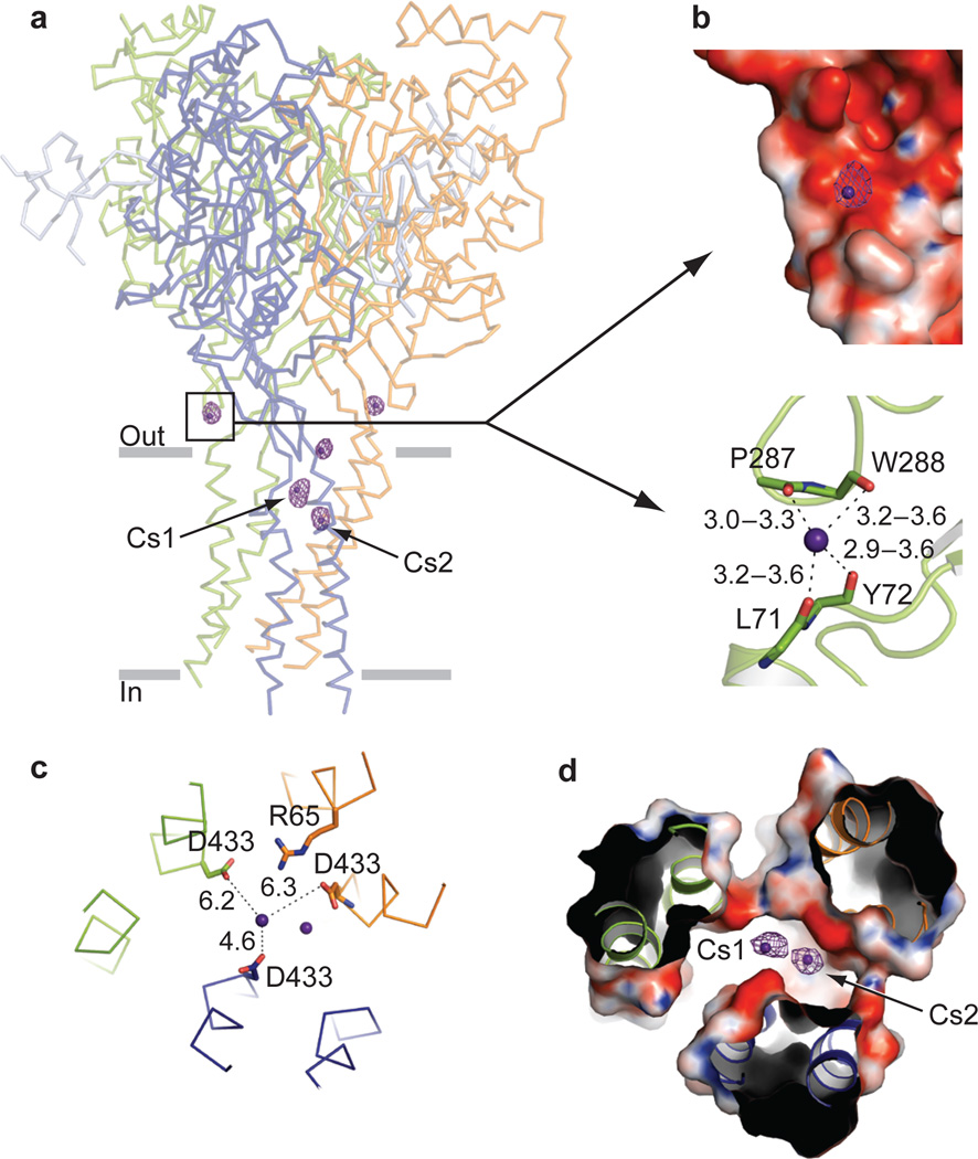
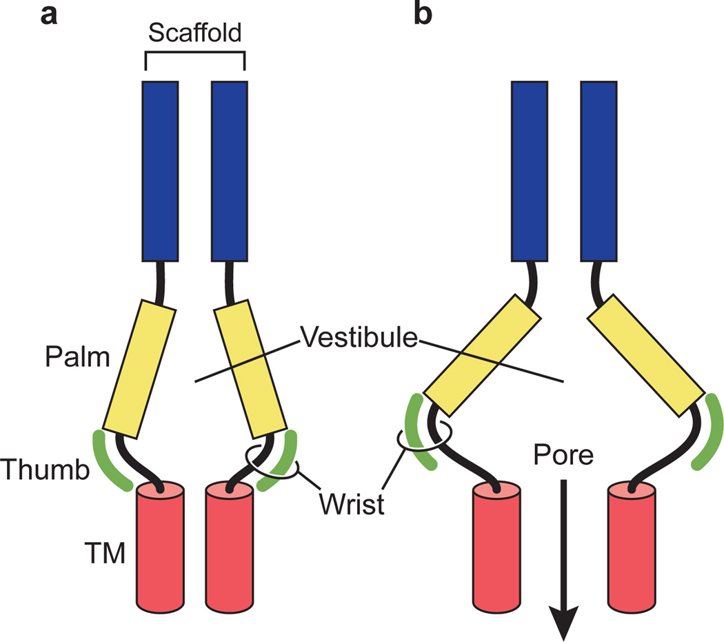
Acid-sensing ion channels (ASICs) are voltage-independent, amiloride-sensitive channels involved in diverse physiological processes ranging from nociception to taste. Despite the importance of ASICs in physiology, we know little about the mechanism of channel activation. Here we show that psalmotoxin activates non-selective and Na(+)-selective currents in chicken ASIC1a at pH 7.25 and 5.5, respectively. Crystal structures of ASIC1a-psalmotoxin complexes map the toxin binding site to the extracellular domain and show how toxin binding triggers an expansion of the extracellular vestibule and stabilization of the open channel pore. At pH 7.25 the pore is approximately 10 Å in diameter, whereas at pH 5.5 the pore is largely hydrophobic and elliptical in cross-section with dimensions of approximately 5 by 7 Å, consistent with a barrier mechanism for ion selectivity. These studies define mechanisms for activation of ASICs, illuminate the basis for dynamic ion selectivity and provide the blueprints for new therapeutic agents.


Figures

Figure 1
Figure 1. PcTx1 activates the chicken ASIC1a Δ13 construct
a, Whole-cell, patch-clamp current traces of activation by steps into pH 5.5 (1), pH 7.25 and 1 µM PcTx1 (2), and pH 5.5 and 1 µM PcTx1 (3). Inset, current trace of step into pH 5.5 and 1 µM PcTx1. b, Structure of low pH Δ13-PcTx1 complex viewed from the extracellular side. c, d, High pH (c) and low pH (d) complexes viewed parallel to the membrane. Each subunit is in a different color and toxin is in solvent-accessible surface representation.
Figure 2
Figure 2. Extensive interactions adhere PcTx1 to the Δ13 ion channel
Close-up views of toxin-binding site of the low pH complex. Dashed lines indicate possible hydrogen bonds.
Figure 3
Figure 3. Conformational changes in the extracellular domain
a, Low pH Δ13-PcTx1 structure is in cartoon representation and colored as in Fig. 2. Black line indicates the axis around which the lower palm domain and the wrist region rotate following superpositions of the desensitized and open state structures. Close-up views of selected regions are boxed. The low pH complex is colored by domain, the high pH complex is orange, and the desensitized state (PDB code: 3HGC) is gray. Approximate position of PcTx1 is indicated by blue dashed lines and the boundaries between adjacent subunits is shown by solid gray lines. Measured Cα distances are between residues Asn 357 (A) and Arg 85 (B) in box 1. In box 2 the distances are between Val 75 Cα atoms on adjacent subunits. b, Close-up view of strands β1 and β12, the β1- β2/β11- β12 linkers and the extracellular boundary of the TM domains from two subunits of the high pH Δ13-PcTx1 complex (orange) and the desensitized state (gray) following superposition of the respective scaffold domains. Inset shows location of close-up view in the context of the entire channel.
Figure 4
Figure 4. Structural rearrangements and ion selectivity of the transmembrane pores
a, b, Comparison of transmembrane domains from the high pH (orange) and desensitized (gray) state structures with TM1 in ribbon and TM2 in cylinder representation. Transmembrane domains are viewed from the extracellular side (a) and parallel to the membrane (b). c, Current/voltage experiment showing that at neutral pH the Δ13-PcTx1 complex forms a non selective cation channel. d, Mapping of solvent accessible pathway along the 3-fold axis shows that the high pH complex has a large, transmembrane pore. The occluded pathway along the 3-fold in the extracellular domain suggests that ions access the pore by way of lateral fenestrations. e, f. A comparison of the transmembrane domains from the low pH complex, where each subunit is in a different color. The desensitized state is gray. TM1 and TM2 segments are in ribbon and cylinder representations, respectively. Transmembrane domains are viewed from the extracellular side (e) and parallel to the membrane (f). g, Current/voltage experiment demonstrating that the ion channel of the low pH Δ13-PcTx1 complex is sodium selective. h, Mapping of a solvent accessible pathway along the pseudo 3-fold axis of the low pH complex shows that it has an asymmetric ion channel pore and a constriction (opposing arrows) halfway across the bilayer. Maps of solvent-accessible pathways (d and h) were generated using the HOLE software (red < 1.4 Å < green < 2.3 Å < purple).
Figure 5
Figure 5. Cs+ binding sites
a, Anomalous difference electron density map contoured at 3.5 σ. The low pH complex is in ribbon representation. Cesium sites in the outer pore region are labeled Cs1 and Cs2. b, Close-up view of the Cs+ site in the wrist region. Top, electrostatic potential contoured from −20kT (red) to +20kT (blue). Bottom, Cs+ coordination by backbone carbonyl oxygens. The main chain is drawn as sticks. c,d, Close-up view of Cs+ sites near Asp 433 shown in ribbon and stick representation (c) and solvent accessible surface colored by electrostatic potential (d) viewed from the extracellular side.
Figure 6
Figure 6. Schematic representation of gating
a, The extracellular vestibule in the closed, desensitized state structure adopts a contracted conformation. b, In the open pore conformation the vestibule occupies an expanded conformation, stabilized by the thumb domain. The wrist region, in turn, couples the conformational changes of the extracellular domain to the transmembrane domains (red cylinder) of the ion channel pore.

References

    1. Gründer S, Chen X. Structure, function, and pharmacology of acid-sensing ion channels (ASICs): focus on ASIC1a. Int J Physiol Pathophysiol Pharmacol. 2010;2:73–94. - PMC - PubMed
    1. Kellenberger S, Schild L. Epithelial sodium channel/degenerin family of ion channels: a variety of functions for a shared structure. Physiol. Rev. 2002;82:735–767. - PubMed
    1. Kashlan OB, Kleyman TR. ENaC structure and function in the wake of a resolved structure of a family member. Am. J. Physiol. Renal. Physiol. 2011;301:F684–F696. - PMC - PubMed
    1. Krishtal OA, Pidoplichko VI. A receptor for protons in the nerve cell membrane. Neuroscience. 1980;5:2325–2327. - PubMed
    1. Waldmann R. Proton-gated cation channels--neuronal acid sensors in the central and peripheral nervous system. Adv Exp Med Biol. 2001;502:293–304. - PubMed

Publication types

MeSH terms