. 2012 Sep;8(9):784-90.
doi: 10.1038/nchembio.1038. Epub 2012 Jul 29.

Peroxidasin forms sulfilimine chemical bonds using hypohalous acids in tissue genesis

Affiliations

Peroxidasin forms sulfilimine chemical bonds using hypohalous acids in tissue genesis

Gautam Bhave et al. Nat Chem Biol. 2012 Sep.

Abstract

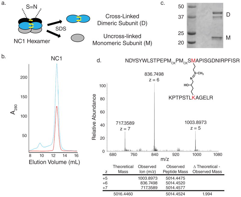
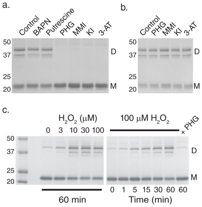
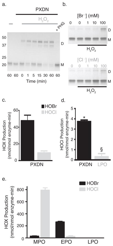
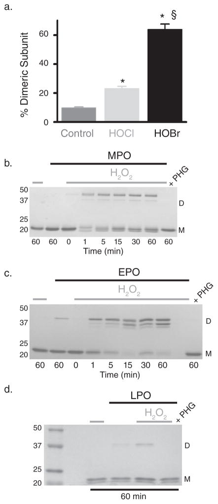
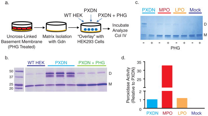
Collagen IV comprises the predominant protein network of basement membranes, a specialized extracellular matrix, which underlie epithelia and endothelia. These networks assemble through oligomerization and covalent crosslinking to endow mechanical strength and shape cell behavior through interactions with cell-surface receptors. A recently discovered sulfilimine (S=N) bond between a methionine sulfur and hydroxylysine nitrogen reinforces the collagen IV network. We demonstrate that peroxidasin, an enzyme found in basement membranes, catalyzes formation of the sulfilimine bond. Drosophila peroxidasin mutants have disorganized collagen IV networks and torn visceral muscle basement membranes, pointing to a critical role for the enzyme in tissue biogenesis. Peroxidasin generates hypohalous acids as reaction intermediates, suggesting a paradoxically anabolic role for these usually destructive oxidants. This work highlights sulfilimine bond formation as what is to our knowledge the first known physiologic function for peroxidasin, a role for hypohalous oxidants in tissue biogenesis, and a possible role for peroxidasin in inflammatory diseases.


Conflict of interest statement

Competing Financial Interests.

The authors have no competing financial interests.

Figures

Figure 1
Figure 1. PFHR-9 cells produce a basement membrane collagen IV network with sulfilimine cross-links
(a) Schematic of collagen IV NC1 hexamer with sulfilimine cross-links bridging the trimer-trimer interface. Upon addition of SDS, the hexamer dissociates into cross-linked dimeric subunits (D) and uncross-linked monomeric subunits (M). (b) Gel filtration chromatography elution profile of PFHR-9 collagen IV NC1 hexamer (blue) and native, purified placental basement membrane NC1 hexamer (red) run successively. (c) SDS-PAGE of the purified NC1 hexamer with cross-linked dimeric (D) and uncross-linked monomeric subunits (M). As seen in placental and mouse Engelbreth-Holm-Swarm (EHS) tumor collagen IV, at least two and occasionally three dimeric subunit bands and one or two monomeric subunit bands were observed. (d) Mass spectrometry of purified PFHR-9 NC1 hexamer revealed a tryptic peptide with a mean observed mass of 5014.4524. The mass of the methionine 93 containing peptide added to the hydroxylysine 211 containing peptide provides a “theoretical” mass of 5016.446. The difference between the theoretical and observed mass of 1.994 represents the loss of two hydrogens upon sulfilimine bond formation in collagen IV. MOX stands for methionine sulfoxide, a common oxidation product of methionine.
Figure 2
Figure 2. A basement membrane peroxidase forms the collagen IV sulfilimine bond
(a) Coomassie blue stained gel after SDS-PAGE of NC1 hexamers isolated from PFHR-9 cells grown in the presence of β-aminopropionitrile (BAPN; 500 μM), putrescine (2.5 mM), phloroglucinol (PHG; 50 μM), methimazole (MMI; 1 mM), 3-aminotriazole (3-AT; 10 mM), and potassium iodide (KI; 10 mM). Collagen IV NC1 hexamer from untreated cells (control) is shown for comparison. Gel is representative of 5 independent experiments. (b) PFHR-9 basement membrane was allowed to form normally, isolated, and treated with phloroglucinol (PHG; 50 μM), methimazole (MMI; 1 mM), potassium iodide (KI; 10 mM), and 3-aminotriazole (3-AT; 10 mM) for 24 hours at 37°C. Collagen IV NC1 hexamer was isolated and underwent SDS-PAGE and Coomassie blue staining to visualize sulfilimine cross-link content. (c) Coomassie blue stained gel after SDS-PAGE of NC1 hexamers after reacting uncross-linked PFHR-9 basement membrane with H2O2 at varying concentrations for 1 hour (left panel) or for varying durations with 100 μM H2O2 (right panel) in 1X PBS (phosphate buffered saline; 150 mM NaCl, 10 mM sodium phosphate, pH 7.4). The gel is representative of 8 independent experiments. D represents NC1 cross-linked dimeric subunits, while M denotes uncross-linked monomeric subunits. Full gel images are provided in Supplementary Fig. 13.
Figure 3
Figure 3. Peroxidasin forms hypohalous acids and sulfilimine bonds in collagen IV
(a) SDS-PAGE of reactions consisting of 16 nM purified human peroxidasin, 500 nM monomeric NC1 hexamer (3 μM potential cross-links), and 10 μM H2O2 in 1X PBS. Control reactions without H2O2 or in the presence of the peroxidase inhibitor, phloroglucinol (PHG; 50 μM), were also conducted. D represents cross-linked dimeric NC1 subunits, while M denotes uncross-linked monomeric subunits. (b) Coomassie stained gel after SDS-PAGE of collagen IV NC1 hexamer is shown to illustrate relative amounts of sulfilimine cross-linked dimeric (D) and uncross-linked monomeric (M) subunits after incubation of uncross-linked PFHR-9 basement membranes in varying buffer halide concentrations (Br or Cl as K+ salt) with or without 1 mM H2O2. (c) Peroxidasin (PXDN) mediated hypohalous acid (HOX) production expressed as nmol hypohalous acid generated per nmol enzyme per minute measured in 1X PBS + 100 μM NaBr. Values represent mean ± s.e.m. (n=3). (d) HOCl production measured directly in 1X PBS without added Br. Values denote mean ± s.e.m. (n=4). PXDN mediated HOCl generation was significantly greater than LPO (*unpaired two-tailed t-test, p < 0.05), while LPO was not statistically different from zero (§ one sample t-test, p = 0.32). (e) Hypohalous acid (HOX) production in nmol hypohalous acid generated per nmol enzyme per minute for myeloperoxidase (MPO), eosinophil peroxidase (EPO), and lactoperoxidase (LPO) in 1X PBS + 100 μM NaBr. Values represent mean ± s.e.m. (n=3). Full gel images are displayed in Supplementary Fig. 14.
Figure 4
Figure 4. Hypohalous acids form collagen IV sulfilimine bonds
(a) 500 nM collagen IV NC1 hexamer (3 μM potential cross-links) was incubated alone (control) or with 5 μM hypochlorous (HOCl) or hypobromous acid (HOBr) for 30 minutes at 37°C. % dimeric subunit (mean ± s.e.m.) as quantified with densitometry of Coomassie stained SDS-PAGE gels (Supplementary Fig. 8) increased significantly with HOCl and HOBr treatment (control n=10, HOCl n=9, HOBr n=6; ANOVA with Tukey’s post-hoc comparison between groups, * = p < 0.05 compared to control, § = p < 0.05 HOCl versus HOBr). (b–d) 16 nM myeloperoxidase (MPO), eosinophil peroxidase (EPO), and lactoperoxidase (LPO) were reacted with 500 nM NC1 hexamer (3 μM potential cross-links) for varying time points in 1X PBS with or without 10 μM H2O2. In the case of LPO, all reactions proceeded for 60 minutes. Collagen IV sulfilimine cross-link content was visualized after SDS-PAGE and Coomassie blue staining of the reactions. Each gel is representative of 3 independent experiments. Complete gel images are provided in Supplementary Fig. 15.
Figure 5
Figure 5. Peroxidasin uniquely cross-links native collagen IV networks
(a) Experimental design of “overlay” experiments. PFHR-9 cells were grown in the presence of phloroglucinol (PHG; 50 μM) to deposit uncross-linked collagen IV networks. The cells were then removed and the basement membrane extracted with 4M guanidine (Gdn) to inactivate endogenous peroxidasin. Human peroxidasin stably transfected cells (PXDN) or untransfected HEK293 cells (WT HEK) were plated on top of the PFHR9 basement membrane, which was subsequently analyzed for collagen IV cross-link content. (b) Collagen IV sulfilimine bond formation in the indicated experimental conditions as demonstrated by stained SDS-PAGE gel. 2 (wildtype HEK cells) or 3 (PXDN transfected with or without PHG) out of 5 independent experiments are displayed. (c) Coomassie stained gel of collagen IV NC1 hexamers isolated from uncross-linked PFHR-9 basement membrane overlaid with HEK293 cells transiently transfected with human peroxidasin cDNA, mouse myeloperoxidase cDNA (MPO), mouse lactoperoxidase cDNA (LPO), or empty vector (Mock). (d) Media from PXDN, MPO, LPO, and mock transfected cells was assayed for peroxidase activity using a tetramethylbenzidine based colorimetric assay. Activity was expressed relative to peroxidasin (A650 of given peroxidase divided by A650 for peroxidasin). Full gel images are displayed in Supplementary Fig. 16.
Figure 6
Figure 6. Peroxidasin is critical for collagen IV and basement membrane integrity
(a) Confocal fluorescence microscopy images of Drosophila anterior midgut using a collagen IV GFP protein trap line (Viking-GFP) to delineate collagen IV distribution. Representative sections from wildtype Pxn +/+ (Pxn +/+), heterozygote Pxn +/− (Pxn+/f07229), and mutant Pxn −/− (Pxnf07229/f07229) flies are shown. Distorted and torn collagen IV networks (arrows) with gross defects (“holes”) in the circumferential muscle layer (asterisks) typified Pxn −/− sections. Scale bar = 10 μm. (b) Immunoblot of collagenase solubilized basement membrane isolated from Drosophila Pxn +/− and Pxn −/− larvae. Pxn −/− mutants demonstrate grossly reduced collagen IV immunoreactivity at 20.4% of wildtype, while Pxn +/− flies have relatively maintained collagen IV NC1 content at 82% of wildtype (Supplementary Fig. 11). Pxn −/− mutants also demonstrated a shift in the % uncross-linked immunoreactivity with 42% of total band density in the uncross-linked form compared to < 9% total band density in Pxn +/− flies (Supplementary Fig. 11).

Similar articles

Cited by

References

    1. Yurchenco PD. Basement membranes: cell scaffoldings and signaling platforms. Cold Spring Harb Perspect Biol. 2011;3:1–27. - PMC - PubMed
    1. Poschl E, et al. Collagen IV is essential for basement membrane stability but dispensable for initiation of its assembly during early development. Development. 2004;131:1619–28. - PubMed
    1. Khoshnoodi J, Pedchenko V, Hudson BG. Mammalian collagen IV. Microsc Res Tech. 2008;71:357–70. - PMC - PubMed
    1. Vanacore R, et al. A sulfilimine bond identified in collagen IV. Science. 2009;325:1230–4. - PMC - PubMed
    1. Pedchenko V, et al. Molecular architecture of the Goodpasture autoantigen in anti-GBM nephritis. N Engl J Med. 2010;363:343–54. - PMC - PubMed

Publication types