. 2012 Oct;110(1):27-36.
doi: 10.1007/s11060-012-0942-0. Epub 2012 Jul 29.

Targeting αV-integrins decreased metastasis and increased survival in a nude rat breast cancer brain metastasis model

Affiliations

Targeting αV-integrins decreased metastasis and increased survival in a nude rat breast cancer brain metastasis model

Y Jeffrey Wu et al. J Neurooncol. 2012 Oct.

Abstract

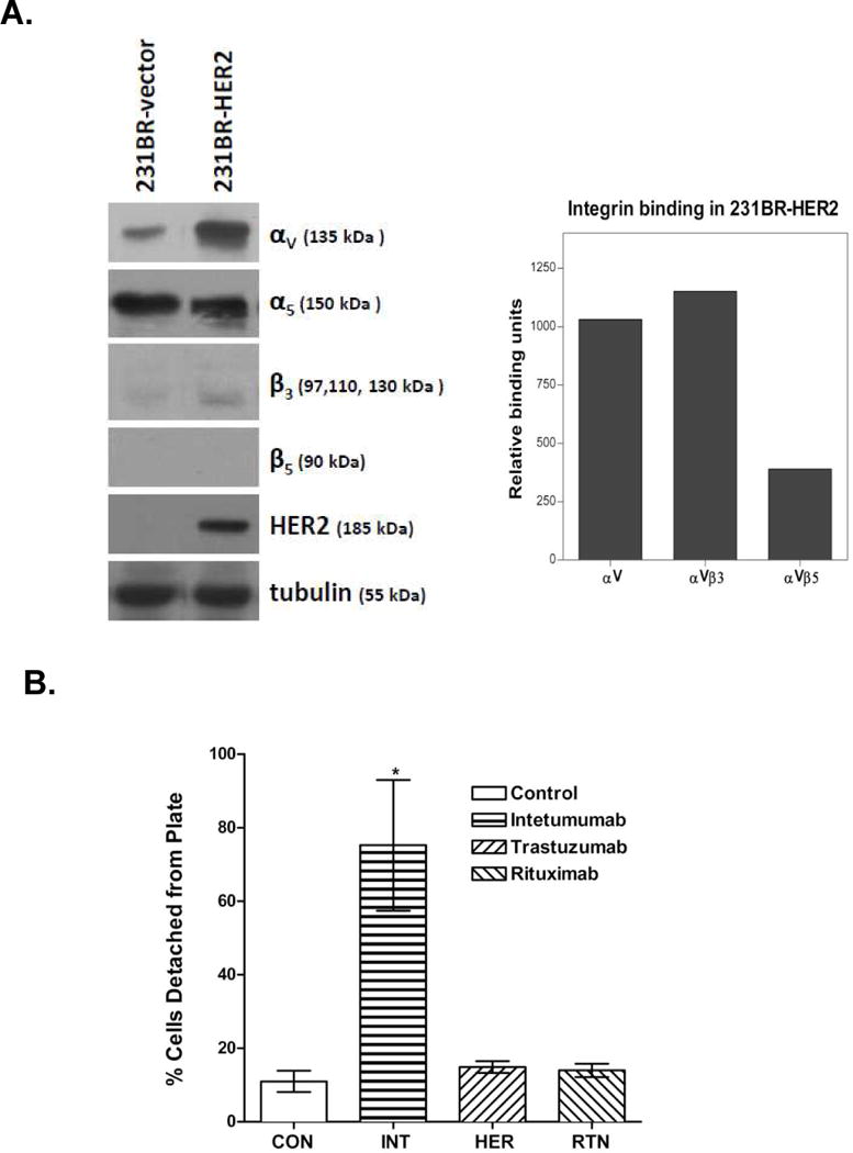
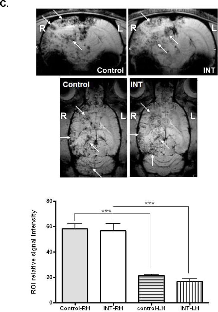
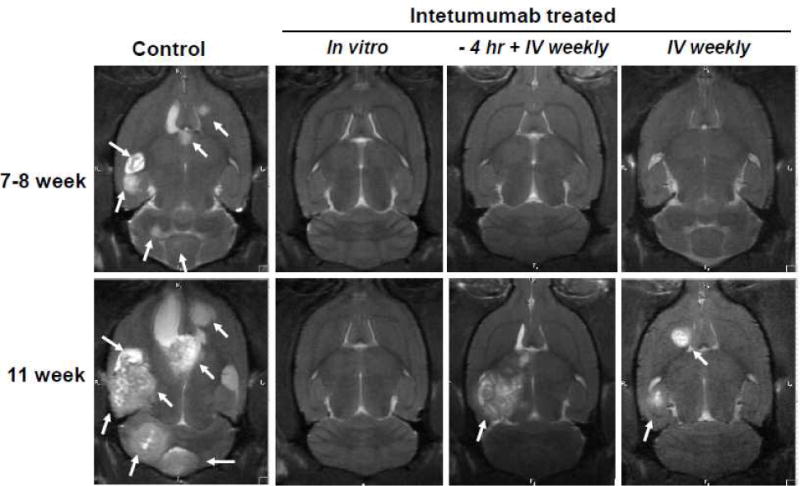
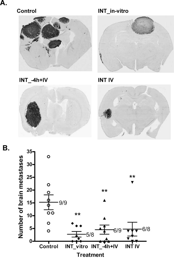
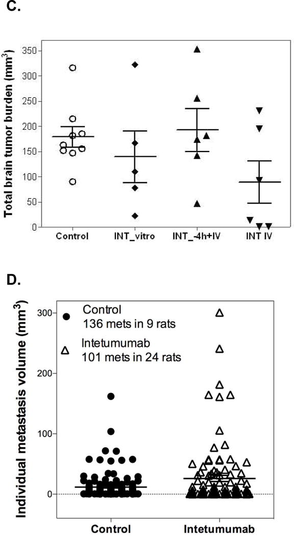
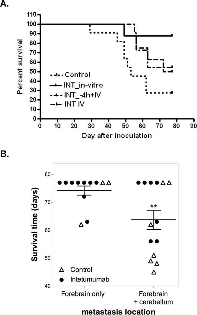
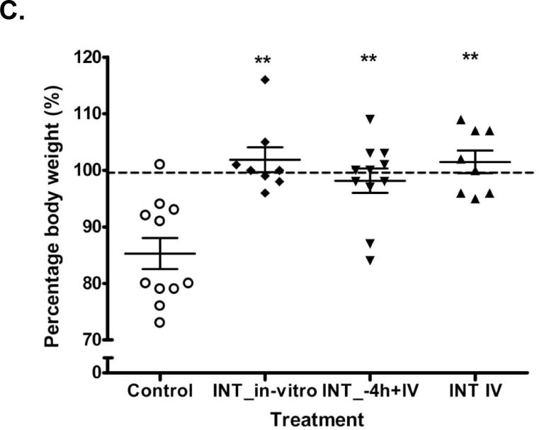
Brain metastases commonly occur in patients with breast, lung and melanoma systemic cancers. The anti-α(V) integrin monoclonal antibody intetumumab binds cell surface proteins important for adhesion, invasion and angiogenesis in the metastatic cascade. The objective of this study was to investigate the anti-metastatic effect of intetumumab in a hematogenous breast cancer brain metastasis model. Female nude rats received intra-carotid infusion of human brain-seeking metastatic breast cancer cells (231BR-HER2) and were randomly assigned into four groups: (1) control; (2) intetumumab mixed with cells in vitro 5 min before infusion without further treatment; (3) intetumumab intravenously 4 h before and weekly after cell infusion; (4) intetumumab intravenously weekly starting 7 days after cell infusion. Brain metastases were detected by magnetic resonance imaging (MRI) and immunohistochemistry. Comparisons were made using the Kruskal-Wallis test and Dunnett's test. Survival times were estimated using Kaplan-Meier analysis. All control rats with brain tissue available for histology (9 of 11 rats) developed multiple brain metastases (median = 14). Intetumumab treatment either in vitro prior to cell infusion or intravenous before or after cell infusion prevented metastasis formation on MRI and decreased the number of metastases on histology (median = 2, p = 0.0055), including 30 % of animals without detectable tumors at the end of the study. The overall survival was improved by intetumumab compared to controls (median 77+ vs. 52 days, p = 0.0277). Our results suggest that breast cancer patients at risk of metastases might benefit from early intetumumab treatment.


Conflict of interest statement

Conflict of Interest: This study was financially sponsored in part by Ortho Biotech Oncology R&D., the manufacturer of the intetumumab antibody. DJM is an employee and stockholder of Ortho Biotech Oncology R&D. The other authors currently have no financial interest and affiliation in this agent, its developer Ortho Biotech Oncology R&D, or its parent company Johnson & Johnson.

Figures

Figure 1
Figure 1. Cellular integrin characterization and effect of Intetumumab treatment on breast cancer cell adhesion in vitro and cell localization in the brain
A) Immunobloting analysis of 231BR-HER2 and their no HER2 expressing matched cell line (231BR-vector) assessed for integrins αv, α5, β3 and β5. HER2 immunoblot was used to confirm the HER2 protein overexpression and anti-tubulin antibody was used as equal loading. In 231BR-HER2 cells, ELISA analysis showed abundant αVβ3 and relatively low αVβ5 integrin dimer. B) Assessment of 23BR-HER2 cell adhesion in vitro. Cells were untreated (CON) or incubated for 1 h with intetumumab (INT, 0.5 mg/mL), trastuzumab (HER, 0.5 mg/mL) or rituximab (RTN, 0.5 mg/mL). Non-adherent cell percentages are indicated as mean ± SD. * indicates p < 0.05. C) Female nude rats received intra-carotid infusion of 231BR-HER2 breast cancer cells labeled with ferumoxides-protamine sulfate and pretreated with either Intetumumab or normal human IgG (0.5 mg/ml) in vitro for 5 min prior to infusion. Coronal and horizontal T2-weighted MRI scans were performed 1 d after cell infusion. Iron is detected as signal dropout on T2-weighted images (arrows). The bar graph indicates quantification of T2* MRI signal intensity of ROI in the right hemisphere (RH) that received intra-arterial cell infusion (on the left in these MRI scans), compared to the left hemisphere (LH). *** indicates p < 0.001.
Figure 2
Figure 2. MRI of the effect of intetumumab on the formation of breast cancer brain metastases
MRI was performed approximately 49–56 and 77 d after intra-carotid cell infusion. One representative rat is shown from each of four randomized treatment groups: 1) Control; 2) INT_in-vitro, cells were pretreated in vitro with intetumumab (0.5 mg/ml) prior to infusion and received no further treatment; 3) INT_-4h+IV, rats received intetumumab (10 mg/kg IV) 4 h prior to cell infusion plus weekly after infusion; 4) Rats received intetumumab (10 mg/kg IV) starting 7 d after cell infusion. The presence of brain lesions (metastases) in MRI was indicated by arrows.
Figure 3
Figure 3. Histological assessment of the effect of intetumumab on breast cancer brain metastasis
After death or sacrifice at 77 d, rat brain sections were assessed for immunohistochemistry against human mitochondria antigen as a marker of human tumor growth. A) One representative rat brain is shown from each group; B) number of brain metastases. Regardless of the route of treatment, intetumumab significantly (** p < 0.01) reduced the number of breast cancer brain metastases in nude rats. The numbers in the plot indicate the number of rats that developed brain metastasis out of total number of rats injected with cancer cells of each treatment. C) Total tumor volume of brain metastases. There was no statistical significance found in tumor volume among treatment groups. Data were presented as dot plot total brain tumor burden of individual rats with mean ± SEM. D) Tumor volume analysis from individual brain metastases in control (n=136 metastases in 9 rats) and intetumumab-treated rats (n=101 metastases in 24 animals). Data are presented as dot plot of individual brain metastasis with mean ± SEM. Intetumumab-treated rats had larger (p=0.0036) average size of individual brain metastases compared to controls by Students t-test.
Figure 4
Figure 4. Effect of intetumumab on survival and body weight of breast cancer brain metastases
A) Kaplan-Meier survival curve: control (dotted line); INT_in-vitro (solid line); INT_-4h+IV (dashed line) and INT_IV (dotted/dashed line). The median survival time was significantly (p=0.0277) improved by intetumumab treatment. B) Brain metastasis location and survival. Data are presented as dot plot of individual rats with mean ± SEM. Lesions within the cerebellum were found more frequently in control rats and were associated with shorter survival (P=0.0058) by Students t-test. C) Plot of percentage of body weight change among the treatment groups. Intetumumab significantly prevented body weight loss compared to control rats. Data were presented as dot plot of individual rat with mean ± SEM. ** indicates p < 0.01.

Similar articles

Cited by

References

    1. Palmieri D, Smith QR, Lockman PR, Bronder J, Gril B, Chambers AF, Weil RJ, Steeg PS. Brain metastases of breast cancer. Breast Dis. 2006;26:139–147. - PubMed
    1. Gril B, Palmieri D, Bronder JL, Herring JM, Vega-Valle E, Feigenbaum L, Liewehr DJ, Steinberg SM, Merino MJ, Rubin SD, Steeg PS. Effect of lapatinib on the outgrowth of metastatic breast cancer cells to the brain. J Natl Cancer Inst. 2008;100:1092–1103. - PMC - PubMed
    1. Palmieri D, Bronder JL, Herring JM, Yoneda T, Weil RJ, Stark AM, Kurek R, Vega-Valle E, Feigenbaum L, Halverson D, Vortmeyer AO, Steinberg SM, Aldape K, Steeg PS. Her-2 overexpression increases the metastatic outgrowth of breast cancer cells in the brain. Cancer Res. 2007;67:4190–4198. - PubMed
    1. Wu YJ, Muldoon LL, Dickey DT, Lewin SJ, Varallyay CG, Neuwelt EA. Cyclophosphamide enhances human tumor growth in nude rat xenografted tumor models. Neoplasia. 2009;11:187–195. - PMC - PubMed
    1. Santarelli JG, Sarkissian V, Hou LC, Veeravagu A, Tse V. Molecular events of brain metastasis. Neurosurg Focus. 2007;22:E1. - PubMed

Publication types

MeSH terms