. 2012 Nov;32(8):1353-65.
doi: 10.1007/s10571-012-9862-y. Epub 2012 Jul 28.

Six commercially available angiotensin II AT1 receptor antibodies are non-specific

Affiliations

Six commercially available angiotensin II AT1 receptor antibodies are non-specific

Julius Benicky et al. Cell Mol Neurobiol. 2012 Nov.

Abstract

Commercially available Angiotensin II AT1 receptor antibodies are widely employed for receptor localization and quantification, but they have not been adequately validated. In this study, six commercially available AT1 receptor antibodies were characterized by established criteria: sc-1173 and sc-579 from Santa Cruz Biotechnology, Inc., AAR-011 from Alomone Labs, Ltd., AB15552 from Millipore, and ab18801 and ab9391 from Abcam. The immunostaining patterns observed were different for every antibody tested, and were unrelated to the presence or absence of AT1 receptors. The antibodies detected a 43 kDa band in western blots, corresponding to the predicted size of the native AT1 receptor. However, identical bands were observed in wild-type mice and in AT1A knock-out mice not expressing the target protein. Moreover, immunoreactivity detected in rat hypothalamic 4B cells not expressing AT1 receptors or transfected with AT1A receptor construct was identical, as revealed by western blotting and immunocytochemistry in cultured 4B cells. Additional prominent immunoreactive bands above and below 43 kDa were observed by western blotting in extracts from tissues of AT1A knock-out and wild-type mice and in 4B cells with or without AT1 receptor expression. In all cases, the patterns of immunoreactivity were independent of the AT1 receptor expression and different for each antibody studied. We conclude that, in our experimental setup, none of the commercially available AT1 receptor antibodies tested met the criteria for specificity and that competitive radioligand binding remains the only reliable approach to study AT1 receptor physiology in the absence of full antibody characterization.


Figures
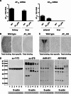
Fig. 1
Fig. 1
Angiotensin II AT1A receptor mRNA, AT1 receptor binding and protein in wild-type, and AT1A knock-out mice (a): expression of mRNAs was studied by RT–PCR in kidney cortex, liver, and adrenal gland of wild-type and AT1A knock-out mice. Gene expression was normalized to the level of 18S rRNA, and represents the mean ± SEM. of three individual determinations. Note that AT1A mRNA is not expressed in kidney cortex and liver of AT1A knock-out mice. AT1B mRNA is not expressed in liver of wild-type or knock-out mice, but it is expressed in adrenal gland of both wild-type and AT1A knock-out mice (b, c): AT1 receptor expression was studied by quantitative autoradiography in sections from kidney cortex (b) and from the hypothalamus (c) containing the PVN (−0.7 mm from bregma), incubated in the presence of 0.25 nM [125I] Sar1–Ile8–angiotensin II as described in “Materials and Methods”. Figures represent a typical result repeated in three different AT1A knock-out and wild-type mice. Note abundant receptor binding in the kidney cortex, predominantly localized to kidney glomeruli, and in the PVN of wild-type mice, completely displaced by incubation with the AT1 receptor-specific antagonist losartan (non-specific binding) and the complete absence of binding in the kidney cortex and PVN of AT1A knockout mice (d): Ang II AT1 receptor protein expression was studied by western blotting. Protein extracts were separated by SDS–PAGE electrophoresis and exposed to four different rabbit polyclonal anti-AT1 receptor antibodies as indicated by catalog number at the top of the picture. Lane 1 liver-WT, 2 liver-AT1AKO, 3 kidney cortex-WT, 4 kidney cortex-AT1AKO, 5 hypothalamus-WT, 6 hypothalamus-AT1AKO. The scale on the left indicates the size in kDa according to the positions of the protein ladder. The expected size of the AT1 receptor is about 43 kDa. Note that in all cases there is no difference between band intensity obtained from tissues from AT1A knock-out and wild-type mice and the presence of many bands not corresponding to the appropriate native AT1 receptor molecular size (43 kDa) with the use of antibodies sc-579, AAR-011, and AB15552. The figure represents a typical experiment repeated two times in individual samples
Fig. 2
Fig. 2
Angiotensin II receptor binding and western blot in control and AT1 receptor-transfected 4B cells (a): expression of AT1A and AT1B mRNA in naïve rat 4B cells. Gene expression was normalized to the level of 18S rRNA and represents the mean ± SEM of three individual determinations. Note that AT1A and AT1B receptor mRNAs were not expressed in rat 4B cells. For comparison, AT1A and AT1B mRNA were quantified in rat kidney and adrenal gland, respectively (b): binding of AT1 receptor- ligand [125I] Sar1–Ang II–AT1A in receptor transfected and control 4B cells. Hypothalamic 4B cells were transiently transfected with a vector containing the entire coding sequence of rat AT1A receptor. Cells transfected with a vector containing the sequence of rat CRH-3′ UTR and non-transfected cells were used as negative controls. The selective AT1 receptor antagonist losartan was used to displace the AT1 receptor-specific binding. Displacement with Ang II was used to determine the non-specific binding. Results are expressed in fmol per 106 cells. Notice the significant AT1 binding expression in AT1A receptor-transfected 4B cells, and the absence of binding in cells transfected with rat CRH-3′ UTR and in non-transfected cells (c): Western blot of protein extracts from AT1A receptor-transfected, CRH-3′ UTR-transfected and non-transfected 4B cells. Protein extracts were separated by SDS–PAGE and exposed to three rabbit polyclonal antibodies against AT1 receptor as indicated by catalog number at the top of the picture. Lane 1 AT1A receptor-transfected 4B cells, 2 CRH-3′ UTR-transfected 4B cells, 3 non-transfected 4B cells. The scale on the left indicates the size in kDa according to the positions of the protein ladder. The expected size of the AT1 receptor is about 43 kDa. Note that AT1 receptor binding is not expressed in control, non-transfected 4B cells, and or in 4B cells transfected with control rat CRH–3′ UTR. Conversely, significant AT1 receptor binding can be detected when 4B cells are transfected with AT1A receptors. Notice that there is no correlation between results obtained by receptor binding and by western blot
Fig. 3
Fig. 3
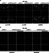
Immunofluorescent staining with AT1 receptor antibodies in non-transfected hypothalamic 4B cells. The cells were stained with sc-1173, sc-579, AB15552, and ab9391 antibodies as described under “Materials and Methods”. Top row staining with AT1 receptor antibodies, middle row nuclear DAPI staining, bottom row merged images. Note that all antibodies reveal significant immunocytochemical staining, and that the staining pattern differs from each of the antibodies studied. Magnification is ×40
Fig. 4
Fig. 4
Immunofluorescent staining with AT1 receptor antibodies in AT1A-transfected hypothalamic 4B cells. a Cells transfected with empty vector (pcDNA3.1) or with rat AT1A receptor (pcDNAI/Amp–rAT1A), and stained with sc-1173, sc-579, and ab9391. Top row antibody staining, middle row: nuclear DAPI staining, bottom row merged images. Note that cells transfected with empty vector express immunoreactivity indistinguishable from that revealed by AT1A receptor transfection. b Cells transfected with empty vector or with AT1A receptors, and stained with AAR-011 or AT15552 antibodies, with or without preabsorption with immunizing peptide. Top row antibody staining, middle row nuclear DAPI staining, bottom row merged images. Note that preabsorption with the immunizing peptide eliminated antibody staining. Magnification is ×40

References

    1. Bader M (2010) Tissue renin–angiotensin-aldosterone systems: targets for pharmacological therapy. Ann Rev Pharmacol Toxicol 50:439–465 - PubMed
    1. Bodei S, Arrighi N, Spano P, Sigala S (2009) Should we be cautious on the use of commercially available antibodies to dopamine receptors? Naunyn Schmiedeberg’s Arch Pharmacol 379:413–415 - PubMed
    1. Burson JM, Aguilera G, Gross KW, Sigmund CD (1994) Differential expression of angiotensin receptor 1A and 1B in mouse. Am J Physiol 267:E260–E267 - PubMed
    1. Chiu AT, Dunscomb J, Kosierowski J, Burton CR, Santomenna LD, Corjay MH, Benfield P (1993) The ligand binding signatures of the rat AT1A, AT1B and the human AT1 receptors are essentially identical. Biochem Biophys Res Commun 197:440–449 - PubMed
    1. Gasc JM, Shanmugam S, Sibony M, Corvol P (1994) Tissue-specific expression of type 1 angiotensin II receptor subtypes. An in situ hybridization study. Hypertension 24:531–537 - PubMed