

Are mechanics different between male and female runners with patellofemoral pain?
- PMID: 22843103
- PMCID: PMC3475738
- DOI: 10.1249/MSS.0b013e3182629215
Are mechanics different between male and female runners with patellofemoral pain?
Abstract
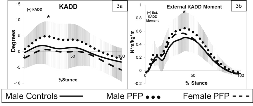
Introduction: Patellofemoral pain (PFP) has often been attributed to abnormal hip and knee mechanics in females. To date, there have been few investigations of the hip and knee mechanics of males with PFP. The purpose of this study was to compare the lower extremity mechanics and alignment of male runners with PFP with healthy male runners and female runners with PFP. We hypothesized that males with PFP would move with greater varus knee mechanics compared with male controls and compared with females with PFP. Furthermore, it was hypothesized that males with PFP would demonstrate greater varus alignment.
Methods: A gait and single-leg squat analysis was conducted on each group (18 runners per group). Measurement of each runner's tibial mechanical axis was also recorded. Motion data were processed using Visual 3D (C-Motion, Bethesda, MD). ANOVAs were used to analyze the data.
Results: Males with PFP ran and squatted in greater peak knee adduction and demonstrated greater peak knee external adduction moment compared with healthy male controls. In addition, males with PFP ran and squatted with less peak hip adduction and greater peak knee adduction compared with females with PFP. The static measure of mechanical axis of the tibial was not different between groups. However, a post hoc analysis revealed that males with PFP ran with greater peak tibial segmental adduction.
Conclusion: Males with PFP demonstrated different mechanics during running and during a single-leg squat compared with females with PFP and with healthy males. Based upon the results of this study, therapies for PFP may need to be sex specific.
Conflict of interest statement
There are no conflicts of interest among any of the authors of this manuscript.
Figures


Similar articles
-
The effect of pain on hip and knee kinematics during running in females with chronic patellofemoral pain.Gait Posture. 2012 Jul;36(3):596-9. doi: 10.1016/j.gaitpost.2012.05.023. Epub 2012 Jun 30. Gait Posture. 2012. PMID: 22749951
-
Prospective evidence for a hip etiology in patellofemoral pain.Med Sci Sports Exerc. 2013 Jun;45(6):1120-4. doi: 10.1249/MSS.0b013e31828249d2. Med Sci Sports Exerc. 2013. PMID: 23274607
-
Are Altered Kinematics in Runners With Patellofemoral Pain Sex Specific?Sports Health. 2022 Nov-Dec;14(6):822-828. doi: 10.1177/19417381221088582. Epub 2022 May 20. Sports Health. 2022. PMID: 35596521 Free PMC article.
-
Gait Retraining With Real-Time Visual Feedback to Treat Patellofemoral Pain in Adult Recreational Runners: A Critically Appraised Topic.J Sport Rehabil. 2019 Nov 7;29(5):675-679. doi: 10.1123/jsr.2019-0094. Print 2020 Jul 1. J Sport Rehabil. 2019. PMID: 31711039 Review.
-
Altered Hip Mechanics and Patellofemoral Pain. A Review of Literature.Ortop Traumatol Rehabil. 2016 May 5;18(3):215-221. doi: 10.5604/15093492.1212855. Ortop Traumatol Rehabil. 2016. PMID: 28157077 Review.
Cited by
-
The current management of patients with patellofemoral pain from the physical therapist's perspective.Ann Jt. 2018 May;3:40. doi: 10.21037/aoj.2018.04.11. Epub 2018 May 14. Ann Jt. 2018. PMID: 31414069 Free PMC article.
-
A 10% Increase in Step Rate Improves Running Kinematics and Clinical Outcomes in Runners With Patellofemoral Pain at 4 Weeks and 3 Months.Am J Sports Med. 2019 Dec;47(14):3406-3413. doi: 10.1177/0363546519879693. Epub 2019 Oct 28. Am J Sports Med. 2019. PMID: 31657964 Free PMC article.
-
Assessment of strength, flexibility, and running mechanics in men with iliotibial band syndrome.J Orthop Sports Phys Ther. 2014 Mar;44(3):217-22. doi: 10.2519/jospt.2014.4991. Epub 2014 Jan 22. J Orthop Sports Phys Ther. 2014. PMID: 24450366 Free PMC article.
-
Relation of Step Length to Magnetic Resonance Imaging-Detected Structural Damage in the Patellofemoral Joint: The Multicenter Osteoarthritis Study.Arthritis Care Res (Hoboken). 2016 Jun;68(6):776-83. doi: 10.1002/acr.22738. Arthritis Care Res (Hoboken). 2016. PMID: 26413842 Free PMC article.
-
Effect of Unanticipated Tasks on Side-Cutting Stability of Lower Extremity with Patellofemoral Pain Syndrome.Sensors (Basel). 2024 Oct 4;24(19):6427. doi: 10.3390/s24196427. Sensors (Basel). 2024. PMID: 39409466 Free PMC article.
References
-
- Barrios J, Davis IS, Higginson JS, Royer T. Lower extremity walking mechanics of young Individuals with asymptomatic varus knee alignment. J Orthop Res. 2009;27(11):1414–1419. - PubMed
-
- Besier TF, Gold GE, Delp SL, Fredericson M, Beaupre GS. The influence of femoral internal and external rotation on cartilage stresses within the patellofemoral joint. J Orthop Res. 2008;26(12):1627–1635. - PubMed
-
- Brinkley J, Stratford P, Lott S, Riddle D. The lower extremity functional scale (LEFS): scale development, measurement properties and clinical application. Phys Ther. 1999;79(4):371–383. - PubMed
-
- Dempster WT, Gabel WC, Felts WJ. The anthropometry of the manual work space for the seated subject. Am. J.Phys. Anthropol. 1959;17:289–317. - PubMed
Publication types
MeSH terms
Grants and funding
LinkOut - more resources
Full Text Sources
Miscellaneous
