

Focal atrophy on MRI and neuropathologic classification of dementia with Lewy bodies
- PMID: 22843258
- PMCID: PMC3413765
- DOI: 10.1212/WNL.0b013e31826357a5
Focal atrophy on MRI and neuropathologic classification of dementia with Lewy bodies
Erratum in
- Neurology. 2012 Sep 4;79(10):1072. Murray, Melissa M [corrected to Murray, Melissa E]
Abstract
Objective: To determine the association between the focal atrophy measures on antemortem MRI and postmortem neuropathologic classification of dementia with Lewy bodies (DLB) using the Third Report of the DLB Consortium criteria.
Methods: We retrospectively identified 56 subjects who underwent antemortem MRI and had Lewy body (LB) pathology at autopsy. Subjects were pathologically classified as high (n = 25), intermediate (n = 22), and low likelihood DLB (n = 9) according to the Third Report of the DLB Consortium criteria. We included 2 additional pathologic comparison groups without LBs: one with low likelihood Alzheimer disease (AD) (control; n = 27) and one with high likelihood AD (n = 33). The associations between MRI-based volumetric measurements and the pathologic classification of DLB were tested with analysis of covariance by adjusting for age, sex, and MRI-to-death interval.
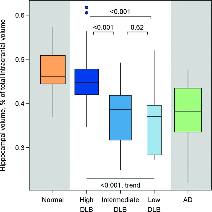
Results: Antemortem hippocampal and amygdalar volumes increased from low to intermediate to high likelihood DLB (p < 0.001, trend test). Smaller hippocampal and amygdalar volumes were associated with higher Braak neurofibrillary tangle stage (p < 0.001). Antemortem dorsal mesopontine gray matter (GM) atrophy was found in those with high likelihood DLB compared with normal control subjects (p = 0.004) and those with AD (p = 0.01). Dorsal mesopontine GM volume decreased from low to intermediate to high likelihood DLB (p = 0.01, trend test).
Conclusion: Antemortem hippocampal and amygdalar volumes increase and dorsal mesopontine GM volumes decrease in patients with low to high likelihood DLB according to the Third Report of the DLB Consortium criteria. Patients with high likelihood DLB typically have normal hippocampal volumes but have atrophy in the dorsal mesopontine GM nuclei.
Figures


References
-
- Schneider JA, Arvanitakis Z, Bang W, Bennett DA. Mixed brain pathologies account for most dementia cases in community-dwelling older persons. Neurology 2007;69:2197–2204 - PubMed
-
- Hansen L, Salmon D, Galasko D, et al. The Lewy body variant of Alzheimer's disease: a clinical and pathologic entity. Neurology 1990;40:1–8 - PubMed
-
- Gomez-Tortosa E, Newell K, Irizarry MC, Albert M, Growdon JH, Hyman BT. Clinical and quantitative pathologic correlates of dementia with Lewy bodies. Neurology 1999;53:1284–1291 - PubMed
-
- Galasko D, Hansen LA, Katzman R, et al. Clinical-neuropathological correlations in Alzheimer's disease and related dementias. Arch Neurol 1994;51:888–895 - PubMed
-
- Fujishiro H, Iseki E, Higashi S, et al. Distribution of cerebral amyloid deposition and its relevance to clinical phenotype in Lewy body dementia. Neurosci Lett 2010;486:19–23 - PubMed
Publication types
MeSH terms
Grants and funding
- R01-AG040042/AG/NIA NIH HHS/United States
- K99-AG037573/AG/NIA NIH HHS/United States
- P50 AG016574/AG/NIA NIH HHS/United States
- AG29550/AG/NIA NIH HHS/United States
- R01 AG011378/AG/NIA NIH HHS/United States
- C06-RR018898/RR/NCRR NIH HHS/United States
- U01-AG 06786/AG/NIA NIH HHS/United States
- R01-AG15866/AG/NIA NIH HHS/United States
- P01-AG017216/AG/NIA NIH HHS/United States
- U01-AG024904-01/AG/NIA NIH HHS/United States
- U01-24904/PHS HHS/United States
- P50-NS072187/NS/NINDS NIH HHS/United States
- R01-AG11378/AG/NIA NIH HHS/United States
- P50-AG16574/AG/NIA NIH HHS/United States
- R21-NS066147/NS/NINDS NIH HHS/United States
- AG32306/AG/NIA NIH HHS/United States
- U24-AG26395/AG/NIA NIH HHS/United States
- AG11378/AG/NIA NIH HHS/United States
- U01-96917/PHS HHS/United States