

Coenzyme Q10 protects retinal cells from apoptosis induced by radiation in vitro and in vivo
- PMID: 22843363
- PMCID: PMC3430426
- DOI: 10.1093/jrr/rrs025
Coenzyme Q10 protects retinal cells from apoptosis induced by radiation in vitro and in vivo
Abstract
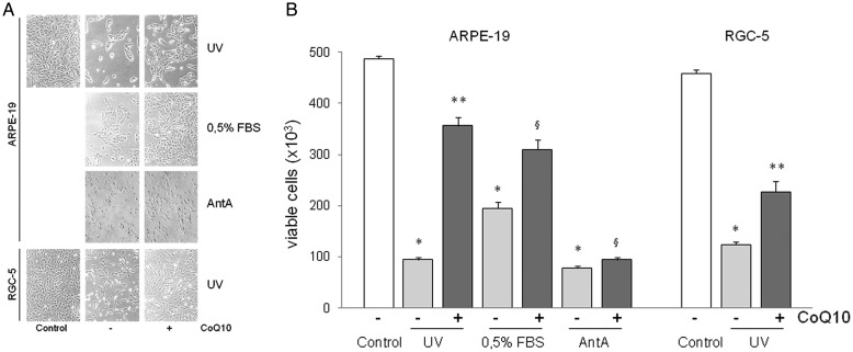
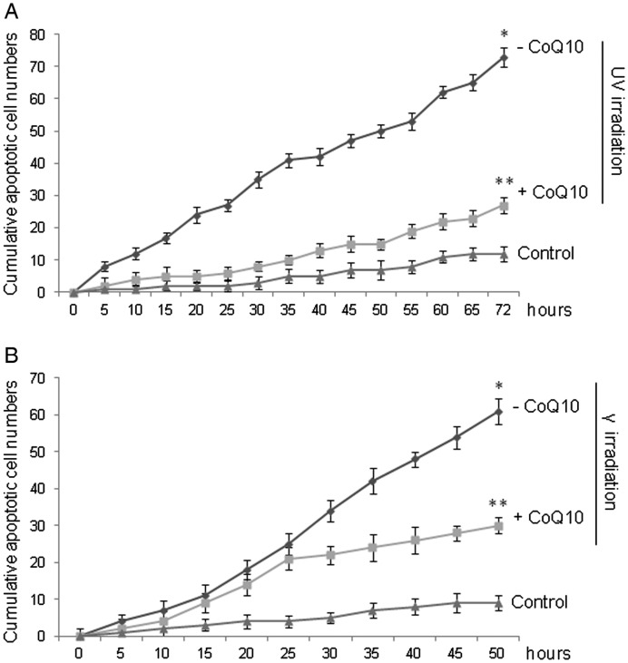
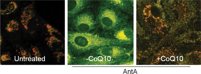
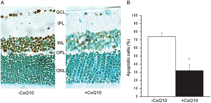
The key pathogenetic event of many retinopathies is apoptosis of retinal cells. Our previous studies have demonstrated that Coenzyme Q10 (CoQ10) prevents apoptosis of corneal keratocytes both in vitro and in vivo, by virtue of its ability to inhibit mitochondrial depolarization, independently of its free radical scavenger role. The aim of this study was to evaluate whether CoQ10 can protect cultured retinal cells and the retinas of rats from radiation-induced apoptosis, if instilled as eye drops in the cornea. In vitro experiments were carried out on cultured ARPE-19 or RGC-5 cells pretreated with CoQ10 before eliciting apoptosis by UV- and γ-radiation, chemical hypoxia (Antimycin A) and serum starvation. Cell viability was evaluated by light microscopy and fluorescence activated cell sorting analysis. Apoptotic events were scored by time-lapse videomicroscopy. Mitochondrial permeability transition was evaluated by JC-1. The anti-apoptotic effectiveness of CoQ10 in retina was also evaluated by an in situ end-labeling assay in Wistar albino rats treated with CoQ10 eye drops prior to UV irradiation of the eye. CoQ10 substantially increased cell viability and lowered retinal cell apoptosis in response both to UV- and γ-radiation and to chemical hypoxia or serum starvation by inhibiting mitochondrion depolarization. In the rat, CoQ10, even when applied as eye drops on the cornea, protected all retina layers from UVR-induced apoptosis. The ability of CoQ10 to protect retinal cells from radiation-induced apoptosis following its instillation on the cornea suggests the possibility for CoQ10 eye drops to become a future therapeutic countermeasure for radiation-induced retinal lesions.
Figures
References
-
- Boulton M, Rózanowska M, Rózanowski B. Retinal photodamage. J Photochem Photobiol B. 2001;64:144–61. - PubMed
-
- Roduit R, Schorderet DF. MAP kinase pathways in UV-induced apoptosis of retinal pigment epithelium ARPE19 cells. Apoptosis. 2008;13:343–53. - PubMed
-
- Brainard GC, Beacham S, Sanford BE, et al. Near ultraviolet radiation elicits visual evoked potentials in children. Clin Neurophysiol. 1999;110:379–83. - PubMed
-
- Roduit R, Schorderet DF. MAP kinase pathways in UV-induced apoptosis of retinal pigment epithelium ARPE19 cells. Apoptosis. 2008;13(3):343–53. - PubMed
