Comparative Study
. 2012 Aug;135(Pt 8):2546-59.
doi: 10.1093/brain/aws175.

Her versus his migraine: multiple sex differences in brain function and structure

Affiliations
Comparative Study

Her versus his migraine: multiple sex differences in brain function and structure

Nasim Maleki et al. Brain. 2012 Aug.

Abstract

Migraine is twice as common in females as in males, but the mechanisms behind this difference are still poorly understood. We used high-field magnetic resonance imaging in male and female age-matched interictal (migraine free) migraineurs and matched healthy controls to determine alterations in brain structure. Female migraineurs had thicker posterior insula and precuneus cortices compared with male migraineurs and healthy controls of both sexes. Furthermore, evaluation of functional responses to heat within the migraine groups indicated concurrent functional differences in male and female migraineurs and a sex-specific pattern of functional connectivity of these two regions with the rest of the brain. The results support the notion of a 'sex phenotype' in migraine and indicate that brains are differentially affected by migraine in females compared with males. Furthermore, the results also support the notion that sex differences involve both brain structure as well as functional circuits, in that emotional circuitry compared with sensory processing appears involved to a greater degree in female than male migraineurs.


Figures

Figure 1
Figure 1
Migraine pain intensity and unpleasantness ratings. A significant difference in the pain unpleasantness scores was observed between the two cohorts (*P < 0.05). The scores are based on a 0–10 subjective scale for migraine pain intensity and pain unpleasantness.
Figure 2
Figure 2
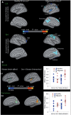
Cortical thickness changes. (A) Significant clusters from vertex-wise cortical thickness comparisons conducted on female versus male healthy subjects (left column) and female versus male migraine patients (right column). Blue–light blue colours represent areas with thicker cortex in female versus male and red–yellow colours represent areas with thicker cortex in male versus male in each of the cohorts. (B) Significant clusters from vertex-wise cortical thickness comparisons conducted on all of the subjects (migraine male and female and healthy control male and female) to determine the main effect (disease) effect and interaction effect (sex × disease). The disease effect (blue–light blue colour map) and sex × disease interaction (red–yellow colour map) are shown for (A) insula and (B) precuneus. The results are Monte Carlo corrected for multiple comparisons. The plots show the average cortical thickness values corresponding to each subject of each cohort that are plotted for each of the clusters representing the main effect of migraine disease. CMF = caudal middle frontal; HC-F = female healthy control; HC-M = male healthy control; IT = inferior temporal; LH = left hemisphere; MIG-F = female migraine; MIG-M = male migraine; PC = precentral gyrus; RH = right hemisphere; RMF = rostral middle frontal; SF = superiorfrontal; SM = supramarginal.
Figure 3
Figure 3
Parahippocampal volume differences. Comparing sex-related volumetric differences between healthy and migraineur subjects revealed significant differences in the parahippocampal gyrus volume as a function of the disease or health state only in males (P < 0.017) but not in females.
Figure 4
Figure 4
Contrast maps for painful heat functional MRI activation. Contrast analysis of the male versus female migraine group in response to the ‘pain threshold +1°C’ stimuli revealed significant (P < 0.05, corrected) differences between the two groups. Caud = caudate; F = female; Hipp = hippocampus; Hypoth = hypothalamus; Ins = insula; L = left; M = male; NAc = nucleus accumbens; PCing = posterior cingulate; Pulv = pulvinar; Put = putamen; R = right; SF = superior frontal; SM = somatosensory cortex; ST = superior temporal.
Figure 5
Figure 5
Conjunction analysis maps for noxious heat response. The maps show common regions of activation for both male and female migraineurs. The active areas are commonly active in both groups but show no significance difference in pain response. Ins = insula; MF = middle frontal; ParaCing = paracingulate; SMA = supplementary motor area; Put = putamen; SM = somatosensory cortex; ST = superior temporal; Thal = thalamus.
Figure 6
Figure 6
Overlap of disease-related and sex-related functional differences. Contrast analysis of the functional differences in female migraineurs versus female healthy control subjects (in green) in response to the ‘pain threshold +1°C’ stimuli revealed significant disease related differences that overlap with sex related differences in migraine patients (in red and blue). Caud = caudate; Hypoth = hypothalamus; Ins = insula; MIG-F = female migraine; MIG-M = male migraine; PCing = paracingulate; PreC = precuneus; Put = putamen; SF = superior frontal.
Figure 7
Figure 7
Functional connectivity contrast maps of the insula and precuneus regions of interest. The insula and precuneus regions of interest were defined for each subject by transforming the cluster of significant cortical thickness differences between migraine and healthy subjects to each subject’s anatomical space. Amyg = amygdala; F = female; M = male; NAc = nucleus accumbens; PoC = postcentral gyrus; Precun = precuneus; SM = somatosensory cortex; TP = temporal pole.

Comment in

Similar articles

Cited by

References

    1. Apkarian AV, Baliki MN, Geha PY. Towards a theory of chronic pain. Progress in neurobiology. 2009;87:81–97. - PMC - PubMed
    1. Apkarian AV, Krauss BR, Fredrickson BE, Szeverenyi NM. Imaging the pain of low back pain: functional magnetic resonance imaging in combination with monitoring subjective pain perception allows the study of clinical pain states. Neurosci Lett. 2001;299:57–60. - PubMed
    1. Becerra L, Morris S, Bazes S, Gostic R, Sherman S, Gostic J, et al. Trigeminal neuropathic pain alters responses in CNS circuits to mechanical (brush) and thermal (cold and heat) stimuli. J Neurosci. 2006;26:10646–57. - PMC - PubMed
    1. Berman SM, Naliboff BD, Suyenobu B, Labus JS, Stains J, Bueller JA, et al. Sex differences in regional brain response to aversive pelvic visceral stimuli. Am J Physiol Regul Integr Comp Physiol. 2006;291:R268–76. - PubMed
    1. Bigal ME, Ashina S, Burstein R, Reed ML, Buse D, Serrano D, et al. Prevalence and characteristics of allodynia in headache sufferers: a population study. Neurology. 2008;70:1525–33. - PMC - PubMed

Publication types