. 2012 Nov 1;21(21):4587-601.
doi: 10.1093/hmg/dds297. Epub 2012 Jul 27.

Enhanced β-secretase processing alters APP axonal transport and leads to axonal defects

Affiliations

Enhanced β-secretase processing alters APP axonal transport and leads to axonal defects

Elizabeth M Rodrigues et al. Hum Mol Genet. .

Abstract

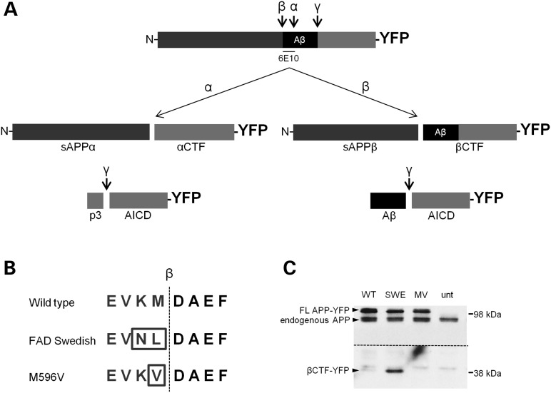
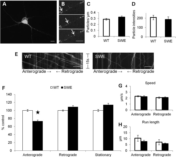
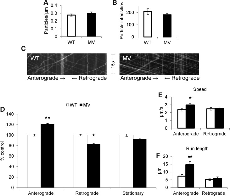
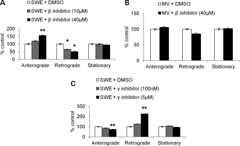
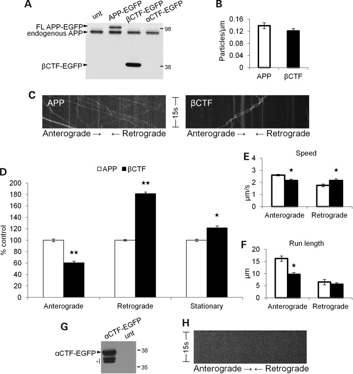
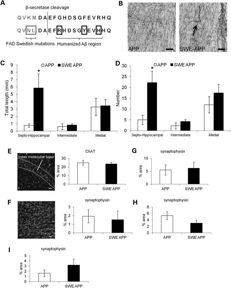
Alzheimer's disease (AD) is a neurodegenerative disease pathologically characterized by amyloid plaques and neurofibrillary tangles in the brain. Before these hallmark features appear, signs of axonal transport defects develop, though the initiating events are not clear. Enhanced amyloidogenic processing of amyloid precursor protein (APP) plays an integral role in AD pathogenesis, and previous work suggests that both the Aβ region and the C-terminal fragments (CTFs) of APP can cause transport defects. However, it remains unknown if APP processing affects the axonal transport of APP itself, and whether increased APP processing is sufficient to promote axonal dystrophy. We tested the hypothesis that β-secretase cleavage site mutations of APP alter APP axonal transport directly. We found that the enhanced β-secretase cleavage reduces the anterograde axonal transport of APP, while inhibited β-cleavage stimulates APP anterograde axonal transport. Transport behavior of APP after treatment with β- or γ-secretase inhibitors suggests that the amount of β-secretase cleaved CTFs (βCTFs) of APP underlies these transport differences. Consistent with these findings, βCTFs have reduced anterograde axonal transport compared with full-length, wild-type APP. Finally, a gene-targeted mouse with familial AD (FAD) Swedish mutations to APP, which enhance the β-cleavage of APP, develops axonal dystrophy in the absence of mutant protein overexpression, amyloid plaque deposition and synaptic degradation. These results suggest that the enhanced β-secretase processing of APP can directly impair the anterograde axonal transport of APP and are sufficient to lead to axonal defects in vivo.


Figures

Figure 1.
Figure 1.
APP-YFP β-secretase cleavage site mutations alter its cleavage. (A) A schematic representation of the APP protein tagged with YFP, with the secretase cleavage sites indicated. Aβ region is indicated in black, and the 6E10 antibody epitope is identified. Cleavage products formed upon α-, β- and γ-secretase cleavage are indicated. (B) Amino acid sequence flanking the β-secretase cleavage site (indicated by dotted line) for WT, SWE and MV APP-YFP. (C) Western blot of SH-SY5Y cells transfected with WT and mutant (SWE and MV) APP-YFP and untransfected (unt) cells as a control. The 6E10 antibody epitope lies in the Aβ region, so it will recognize full-length APP-YFP and βCTF-YFP, but not the αCTF-YFP. Top arrowheads identify full-length APP-YFP and full-length endogenous APP bands, and bottom arrowhead identifies βCTF-YFP bands. Both YFP bands are absent in the untransfected control. Above the dotted line is the membrane at a low exposure. Below the dotted line is the same membrane at a higher exposure to reveal the βCTF-YFP bands. This higher exposure also reveals a set of non-specific bands present in all samples that lies immediately above the βCTF-YFP bands.
Figure 2.
Figure 2.
FAD Swedish mutations reduce APP anterograde axonal transport. (A) Confocal image of a mouse hippocampal neuron transfected with SWE APP-YFP. Scale bar = 10 µm (B) Fluorescent SWE APP-YFP particles in an axon. Arrows follow a single particle traveling in the anterograde direction across chronological frames (top to bottom) from a movie used in data analysis. Scale bar = 10 µm. (CH) WT APP-YFP (white) n = 40 axons, 1009 particles and SWE APP-YFP (black) n = 40 axons, 1129 particles. (C) The mean number of fluorescent particles per length of transfected mouse hippocampal axon. (D) The mean fluorescent intensities of axonal particles. (E) Representative kymographs for WT and FAD Swedish APP-YFP axonal transport from 15s, 10 Hz movies. Right or left descending particles represent anterograde or retrograde moving vesicles, respectively. Vertical lines represent stationary particles. (F) Percentage of anterograde (P = 0.017), retrograde and stationary SWE APP-YFP particles compared with WT. Average segmental velocities (G) and segmental run lengths (H) for anterograde and retrograde APP-YFP particles obtained from kymograph analyses. (Student's t-tests, *P < 0.05.
Figure 3.
Figure 3.
MV mutation stimulates APP anterograde axonal transport. (AF) WT APP-YFP (white) n = 44 axons, 1172 particles, and MV APP-YFP (black) n = 52 axons, 1577 particles. (A) The mean number of fluorescent particles per length of transfected mouse hippocampal axon. (B) The mean fluorescent intensities of axonal particles. (C) Representative kymographs for WT and MV APP-YFP axonal transport. (D) Percentage of anterograde (P = 0.007), retrograde (P = 0.045) and stationary MV APP-YFP particles compared with WT. Average segmental velocities (E) and segmental run lengths (F) for anterograde (P = 0.026 and P = 0.003, respectively) and retrograde APP-YFP particles obtained from kymograph analyses (Student's t-tests, *P < 0.05, **P < 0.01).
Figure 4.
Figure 4.
Pharmacological inhibition of β- and γ-secretase activity alters APP axonal transport. (A) Percentage of anterograde, retrograde and stationary SWE APP-YFP particles treated with 10 µm β-secretase inhibitor (gray, n = 25 axons, 598 particles) or 40 µm β-secretase inhibitor (black, n = 22 axons, 445 particles) compared with DMSO (white, n = 25 axons and 697 particles for 10 µm data set, n = 18 axons and 234 particles for 40 µm data set). Anterograde axonal transport significantly increased with 40 µm β-secretase inhibitor (P = 0.006), while retrograde transport decreased with both 10 and 40 µm inhibitor (P = 0.018 and P = 0.019, respectively). (B) Percentage of anterograde, retrograde and stationary MV APP-YFP particles treated with 40 µm β-secretase inhibitor (black, n = 33 axons, 309 particles) compared with DMSO (white, n = 29 axons, 373 particles). (C) Percentage of anterograde (P = 0.002), retrograde (P = 0.001) and stationary SWE APP-YFP particles treated with 100 nm (n = 49 axons, 1499 particles) or 5 µm γ-secretase inhibitor (black, n = 22 axons, 674 particles) compared with DMSO (white, n = 35 axons and 995 particles for 100 nm data set, n = 12 axons and 322 particles for 5 µm data set) (Student's t tests, *P < 0.05, **P < 0.01).
Figure 5.
Figure 5.
βCTFs have reduced anterograde axonal transport. (A) Western blot of SH-SY5Y cells transfected with WT APP-EGFP, βCTF-EGFP, αCTF-EGFP and untransfected (unt) cells as a control. The 6E10 antibody epitope lies in the Aβ region, so it will recognize full-length APP-EGFP and βCTF-EGFP, but not αCTF-EGFP. Top arrowhead identifies the full-length APP-EGFP band present only in APP-EGFP transfected cells, and bottom arrowhead identifies the βCTF-EGFP band that is absent in αCTF-EGFP transfected cells. (BF) WT APP-EGFP (white) n = 52 axons, 658 particles, βCTF-EGFP (black) n = 38 axons, 448 particles. (B) The mean number of fluorescent particles per length of transfected mouse hippocampal axon. (C) Representative kymographs for WT APP-EGFP and βCTF-EGFP axonal transport. (D) Percentage of anterograde (P < 0.001), retrograde (P = 0.003) and stationary (P = 0.024) particles for βCTF-EGFP compared with WT APP-EGFP. Segmental velocities (E) and run lengths (F) for anterograde (P = 0.001 and P < 0.001, respectively) and retrograde (P = 0.025) particles obtained from kymograph analyses. (G) Western blot of cells transfected with αCTF-EGFP and the untransfected control, probed with a C-terminal APP antibody. Below αCTF-EGFP product lies two bands of unidentified/degradation products (indicated by asterisk), which were previously reported with the βCTF-EGFP plasmid from which this αCTF-EGFP plasmid was constructed (85). (H) Representative kymograph for αCTF-EGFP showing no axonal transport (Student's t-tests, *P < 0.05, **P < 0.01).
Figure 6.
Figure 6.
Axonal defects in FAD Swedish APP gene-targeted mice. (A) WT (top) and mutant (bottom) amino acid sequences near the β-secretase cleavage site for mouse APP. Mutated amino acids are boxed in the bottom row. (B) ChAT immunohistochemistry in the septohippocampal subregion of the septal nucleus in the basal forebrain. Dilated axon (arrow) shown in the FAD Swedish APP brain. Scale bar = 30 µm. (C) The average length and (D) the average number of dilated axons with diameter over 1.5 µm in septohippocampal, intermediate and medial subregions of the septal nucleus at 12 months [WT, n = 4 (white); APP, n = 4 (black)]. (E) (Left) Example image of ChAT staining in the dentate gyrus of the hippocampus. (Right) The average percent area labeled by ChAT in inner molecular layer of the dentate gyrus in 18 m mice [WT, n = 4 (white); APP, n = 4 (black)]. (F) (Left) Example image of synaptophysin staining in the dentate gyrus. (Right) The average percent area labeled by synaptophysin in the inner molecular layer of dentate gyrus in 6 m mice (WT, n = 4; APP, n = 3) and (G) 18 m mice (WT, n = 3; APP, n = 4). The average percent area labeled by synaptophysin in the hippocampal region (H) CA1 in 18 m mice (WT, n = 3; APP, n = 4) and (I) CA3 in 24 m mice (WT, n = 3; APP, n = 3) (Mann–Whitney rank-sum test, *α < 0.05).

Similar articles

Cited by

References

    1. Papp H., Pakaski M., Kasa P. Presenilin-1 and the amyloid precursor protein are transported bidirectionally in the sciatic nerve of adult rat. Neurochem. Int. 2002;41:429–435. - PubMed
    1. Gunawardena S., Goldstein L.S. Disruption of axonal transport and neuronal viability by amyloid precursor protein mutations in Drosophila. Neuron. 2001;32:389–401. - PubMed
    1. Yamazaki T., Selkoe D.J., Koo E.H. Trafficking of cell surface beta-amyloid precursor protein: retrograde and transcytotic transport in cultured neurons. J. Cell Biol. 1995;129:431–442. - PMC - PubMed
    1. Sisodia S.S., Koo E.H., Hoffman P.N., Perry G., Price D.L. Identification and transport of full-length amyloid precursor proteins in rat peripheral nervous system. J. Neurosci. 1993;13:3136–3142. - PMC - PubMed
    1. Koo E.H., Sisodia S.S., Archer D.R., Martin L.J., Weidemann A., Beyreuther K., Fischer P., Masters C.L., Price D.L. Precursor of amyloid protein in Alzheimer disease undergoes fast anterograde axonal transport. Proc. Natl Acad. Sci. USA. 1990;87:1561–1565. - PMC - PubMed

Publication types

MeSH terms

Substances