. 2012 Oct;78(19):7003-11.
doi: 10.1128/AEM.02018-12. Epub 2012 Jul 27.

Construction and application of a luxABCDE reporter system for real-time monitoring of Enterococcus faecalis gene expression and growth

Affiliations

Construction and application of a luxABCDE reporter system for real-time monitoring of Enterococcus faecalis gene expression and growth

Sabina Leanti La Rosa et al. Appl Environ Microbiol. 2012 Oct.

Abstract

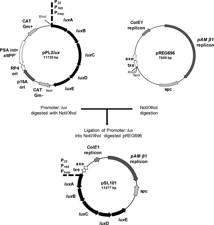
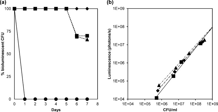
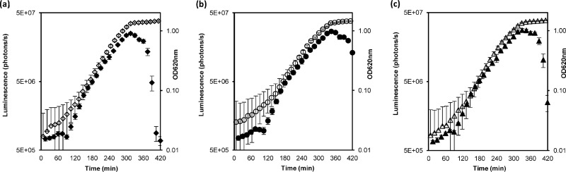
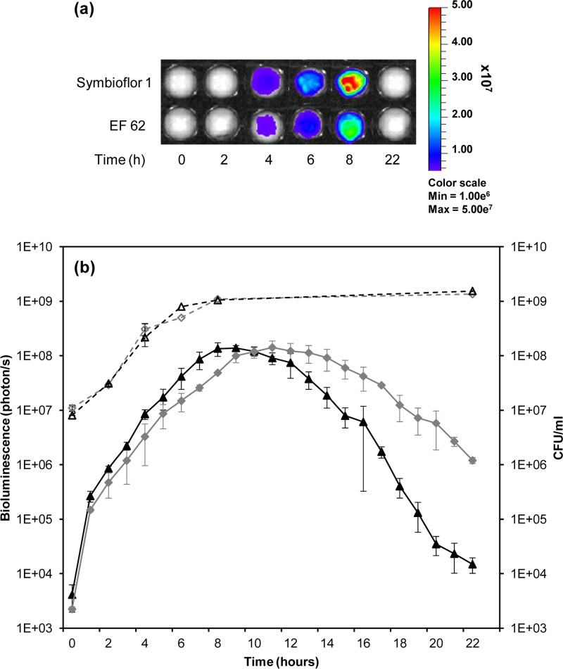
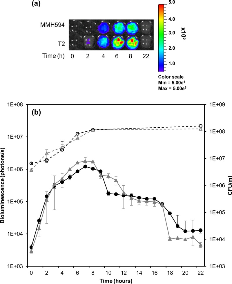
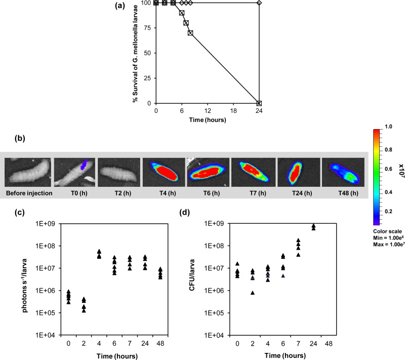
The present work describes the construction of a novel molecular tool for luciferase-based bioluminescence (BL) tagging of Enterococcus faecalis. To this end, a vector (pSL101) and its derivatives conferring a genetically encoded bioluminescent phenotype on all tested strains of E. faecalis were constructed. pSL101 harbors the luxABCDE operon from pPL2lux and the pREG696 broad-host-range replicon and axe-txe toxin-antitoxin cassette, providing segregational stability for long-term plasmid persistence in the absence of antibiotic selection. The bioluminescent signals obtained from three highly expressed promoters correlated linearly (R(2) > 0.98) with the viable-cell count. We employed lux-tagged E. faecalis strains to monitor growth in real time in milk and urine in vitro. Furthermore, bioluminescence imaging (BLI) was used to visualize the magnitude of the bacterial burden during infection in the Galleria mellonella model system. To our knowledge, pSL101 is the first substrate addition-independent reporter system developed for BLI of E. faecalis and an efficient tool for spatiotemporal tracking of bacterial growth and quantitative determination of promoter activity in real time, noninvasively, in infection model systems.


Figures

Fig 1
Fig 1
Strategy for the construction of vector pSL101. The promoter of interest was cloned in pPL2lux, and subsequently, the promoter::lux NotI/XhoI cassettes were subcloned in pREG696, producing pSL101 and derivatives. See Materials and Methods for details.
Fig 2
Fig 2
(a) In vitro plasmid stability of pSL101 derivative constructs in E. faecalis. ◆, SL03 (MMH594::pSL101P32); ■, SL04 (MMH594::pSL101P16S); ▲, SL05 (MMH594::pSL101Phelp); and ●, SL02 (MMH594::pIL252luxPhelp). The strains were subcultured over 7 days without selective pressure at 37°C in GM17, and dilutions were plated on the appropriate antibiotic. The stability of the plasmid is expressed as a percentage of bioluminescent colonies on a nonselective plate compared to that on a selective plate. (b) Correlation between bioluminescence emission and viable-cell counts (CFU) in E. faecalis. ◆, SL03 (MMH594::pSL101P32) (R2 = 0.993); ■, SL04 (MMH594::pSL101P16S) (R2 = 0.995); ▲, SL05 (MMH594::pSL101Phelp) (R2 = 0.990). Samples from the mid-logarithmic phase were serially diluted, and bioluminescence was determined. The last two dilutions were plated on selective media to determine viable bacterial counts. The linear regression coefficient (R2) shows a linear relationship between luminescence emission and the number of CFU per ml. Bioluminescence values are expressed as photons per second. The values represent the averages of two biological replicates.
Fig 3
Fig 3
Bioluminescence during growth in GM17 at 37°C. The closed symbols indicate the bioluminescent signal in photons per second, while growth is represented by open symbols. (a) SL03 (MMH594::pSL101P32). (b) SL04 (MMH594::pSL101P16S). (c) SL05 (MMH594::pSL101Phelp). Growth was measured as the optical density at 620 nm. The values represent the averages of three independent experiments ± standard deviations (SD).
Fig 4
Fig 4
(a) In vitro bioluminescence monitoring during growth at 37°C in Nestle NAN Infant Milk Formula. The color scale indicates bioluminescence signal intensity in photons per second (minimum, 1.0e6; maximum, 5.0e7). (b) Comparison between bioluminescence emission (photons per second) and number of CFU during growth in milk. Bioluminescence is indicated by black closed triangles for SL06 (Symbioflor 1::pSL101P16S) and gray closed diamonds for SL08 (EF62::pSL101P16S). Growth is indicated by open symbols and expressed as CFU per milliliter. The values represent averages of two independent experiments ± SD.
Fig 5
Fig 5
(a) In vitro bioluminescence monitoring during growth at 37°C in urine. The color scale indicates bioluminescence signal intensity in photons per second (minimum, 5.0e4; maximum, 5.0e5). (b) Comparison between photon emission and number of CFU during growth in urine. Bioluminescence is indicated by black closed circles for E. faecalis SL04 (MMH594::pSL101P16S) and gray closed triangles for SL09 (T2::pSL101P16S). Growth is indicated by open symbols and expressed as CFU per milliliter. Bioluminescence was measured as photons per second. The values represent averages of the data from two independent experiments ± SD.
Fig 6
Fig 6
(a) Killing of G. mellonella larvae by E. faecalis MMH594 (□) and SL04 (MMH594::pSL101P16S) (×). The larvae were infected with approximately 4 × 10⁶ bacteria. As a control, 10 larvae were injected with 10 μl 0.9% saline solution (♢). The data were obtained from two independent experiments. (b) Monitoring of E. faecalis SL04 colonization in G. mellonella by bioluminescence imaging of the intact insects. T, time. (c) Light emission detected over 48 h corresponding to the growth of E. faecalis SL04. (d) CFU of E. faecalis SL04 (MMH594::pSL101P16S) from homogenized G. mellonella larvae.

Similar articles

Cited by

References

    1. Andreu N, et al. 2010. Optimisation of bioluminescent reporters for use with mycobacteria. PLoS One 5:e10777 doi:10.1371/journal.pone.0010777 - DOI - PMC - PubMed
    1. Arends K, Schiwon K, Sakinc T, Hubner J, Grohmann E. 2012. Green fluorescent protein-labeled monitoring tool to quantify conjugative plasmid transfer between Gram-positive and Gram-negative bacteria. Appl. Environ. Microbiol. 78:895–899 - PMC - PubMed
    1. Bachmann H, Santos F, Kleerebezem M, van Hylckama Vlieg JE. 2007. Luciferase detection during stationary phase in Lactococcus lactis. Appl. Environ. Microbiol. 73:4704–4706 - PMC - PubMed
    1. Branchini BR, et al. 2007. Thermostable red and green light-producing firefly luciferase mutants for bioluminescent reporter applications. Anal. Biochem. 361:253–262 - PubMed
    1. Brede DA, Snipen LG, Ussery DW, Nederbragt AJ, Nes IF. 2011. Complete genome sequence of the commensal Enterococcus faecalis 62, isolated from a healthy Norwegian infant. J. Bacteriol. 193:2377–2378 - PMC - PubMed

Publication types

LinkOut - more resources