

Amelioration of radiation-induced hematopoietic and gastrointestinal damage by Ex-RAD(R) in mice
- PMID: 22843617
- PMCID: PMC3393340
- DOI: 10.1093/jrr/rrs001
Amelioration of radiation-induced hematopoietic and gastrointestinal damage by Ex-RAD(R) in mice
Abstract
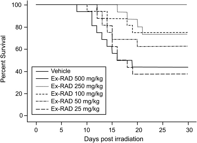
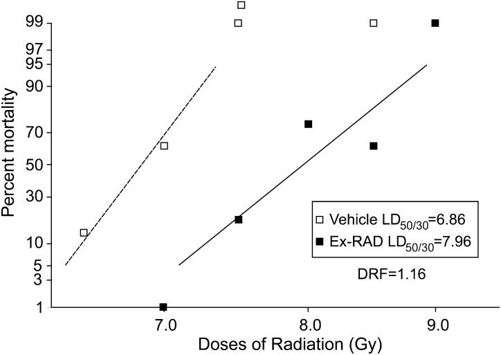
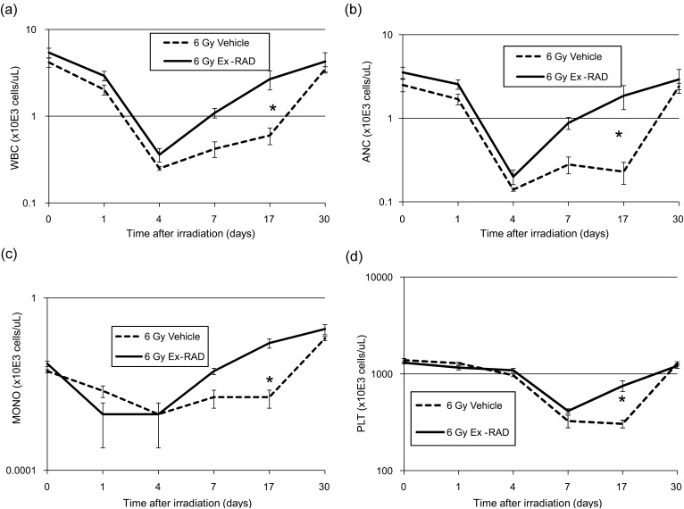
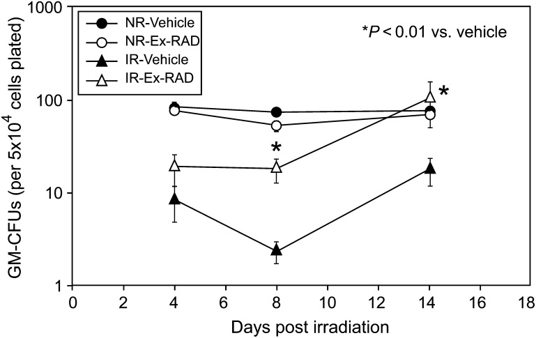
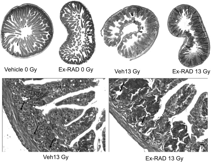
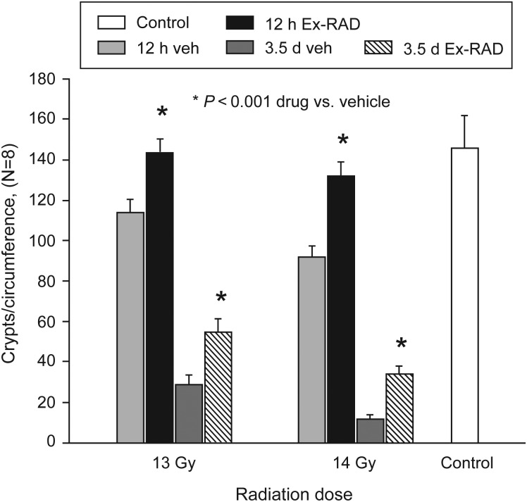
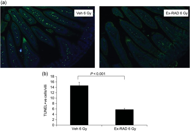
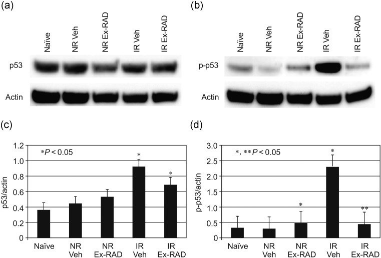
The aim of the present study was to assess recovery from hematopoietic and gastrointestinal damage by Ex-RAD(®), also known as ON01210.Na (4-carboxystyryl-4-chlorobenzylsulfone, sodium salt), after total body radiation. In our previous study, we reported that Ex-RAD, a small-molecule radioprotectant, enhances survival of mice exposed to gamma radiation, and prevents radiation-induced apoptosis as measured by the inhibition of radiation-induced protein 53 (p53) expression in cultured cells. We have expanded this study to determine best effective dose, dose-reduction factor (DRF), hematological and gastrointestinal protection, and in vivo inhibition of p53 signaling. A total of 500 mg/kg of Ex-RAD administered at 24 h and 15 min before radiation resulted in a DRF of 1.16. Ex-RAD ameliorated radiation-induced hematopoietic damage as monitored by the accelerated recovery of peripheral blood cells, and protection of granulocyte macrophage colony-forming units (GM-CFU) in bone marrow. Western blot analysis on spleen indicated that Ex-RAD treatment inhibited p53 phosphorylation. Ex-RAD treatment reduces terminal deoxynucleotidyl transferase mediated dUTP nick end labeling assay (TUNEL)-positive cells in jejunum compared with vehicle-treated mice after radiation injury. Finally, Ex-RAD preserved intestinal crypt cells compared with the vehicle control at 13 and 14 Gy. The results demonstrated that Ex-RAD ameliorates radiation-induced peripheral blood cell depletion, promotes bone marrow recovery, reduces p53 signaling in spleen and protects intestine from radiation injury.
Figures
Similar articles
-
Pharmacological Modulation of Radiation Damage. Does It Exist a Chance for Other Substances than Hematopoietic Growth Factors and Cytokines?Int J Mol Sci. 2017 Jun 28;18(7):1385. doi: 10.3390/ijms18071385. Int J Mol Sci. 2017. PMID: 28657605 Free PMC article. Review.
-
Administration of ON 01210.Na after exposure to ionizing radiation protects bone marrow cells by attenuating DNA damage response.Radiat Oncol. 2012 Jan 20;7:6. doi: 10.1186/1748-717X-7-6. Radiat Oncol. 2012. PMID: 22264334 Free PMC article.
-
Radiation protection by a new chemical entity, Ex-Rad: efficacy and mechanisms.Radiat Res. 2009 Feb;171(2):173-9. doi: 10.1667/RR1367.1. Radiat Res. 2009. PMID: 19267542
-
α-Tocopherol succinate protects mice against radiation-induced gastrointestinal injury.Radiat Res. 2012 Feb;177(2):133-45. doi: 10.1667/rr2627.1. Epub 2011 Oct 20. Radiat Res. 2012. PMID: 22013885
-
Radiation protection by Ex-RAD: a systematic review.Environ Sci Pollut Res Int. 2020 Sep;27(27):33592-33600. doi: 10.1007/s11356-020-09618-y. Epub 2020 Jun 25. Environ Sci Pollut Res Int. 2020. PMID: 32583118
Cited by
-
Synthesis and radioprotective effects of novel benzyl naphthyl sulfoxide (sulfone) derivatives transformed from Ex-RAD.Medchemcomm. 2018 Feb 19;9(4):625-631. doi: 10.1039/c7md00573c. eCollection 2018 Apr 1. Medchemcomm. 2018. PMID: 30108953 Free PMC article.
-
Protective effects of celastrol against γ irradiation-induced oxidative stress in human umbilical vein endothelial cells.Exp Ther Med. 2018 Aug;16(2):685-694. doi: 10.3892/etm.2018.6270. Epub 2018 Jun 7. Exp Ther Med. 2018. PMID: 30116323 Free PMC article.
-
Substituted Piperazines as Novel Potential Radioprotective Agents.Molecules. 2020 Jan 25;25(3):532. doi: 10.3390/molecules25030532. Molecules. 2020. PMID: 31991816 Free PMC article.
-
Pharmacological Modulation of Radiation Damage. Does It Exist a Chance for Other Substances than Hematopoietic Growth Factors and Cytokines?Int J Mol Sci. 2017 Jun 28;18(7):1385. doi: 10.3390/ijms18071385. Int J Mol Sci. 2017. PMID: 28657605 Free PMC article. Review.
-
Mitigation of total body irradiation-induced mortality and hematopoietic injury of mice by a thrombopoietin mimetic (JNJ-26366821).Sci Rep. 2022 Mar 3;12(1):3485. doi: 10.1038/s41598-022-07426-7. Sci Rep. 2022. PMID: 35241733 Free PMC article.
References
-
- Reeves GI. Radiation injuries. Crit Care Clin. 1999;15:457–473. - PubMed
-
- Fliedner TM, Nothdurft W, Heit H. Biological factors affecting the occurrence of radiation syndromes. In: Broerse JJ, MacVittie TJ, editors. Response of Different Species to Total Body Irradiation. Leiden: Martinas Nijhoff Publishers; 1984. pp. 209–219.
-
- Hendry JH, Feng-Tong Y. Response of bone marrow to low LET irradiation. In: Hendry JH, Lord BI, editors. Radiation Toxicology: Bone Marrow and Leukemia. London: Taylor and Francis; 1995. pp. 91–116.
-
- Mauch P, Constine L, Greenberger J, et al. Hematopoietic stem cell compartment: acute and late effects of radiation therapy and chemotherapy. Int J Radiat Oncol Biol Physet. 1995;31:1319–39. - PubMed
-
- Neta R, Oppenheim JJ, Douches SD. Interdependence of the radioprotective effects of human recombinant interleukin 1 alpha, tumor necrosis factor alpha, granulocyte colony-stimulating factor, and murine recombinant granulocyte-macrophage colony-stimulating factor. J Immunol. 1988;140:108–11. - PubMed
Publication types
MeSH terms
Substances
LinkOut - more resources
Full Text Sources
Research Materials
Miscellaneous
