. 2012 Sep 14;287(38):31973-82.
doi: 10.1074/jbc.M112.348565. Epub 2012 Jul 26.

The arginine of the DRY motif in transmembrane segment III functions as a balancing micro-switch in the activation of the β2-adrenergic receptor

Affiliations

The arginine of the DRY motif in transmembrane segment III functions as a balancing micro-switch in the activation of the β2-adrenergic receptor

Louise Valentin-Hansen et al. J Biol Chem. .

Abstract

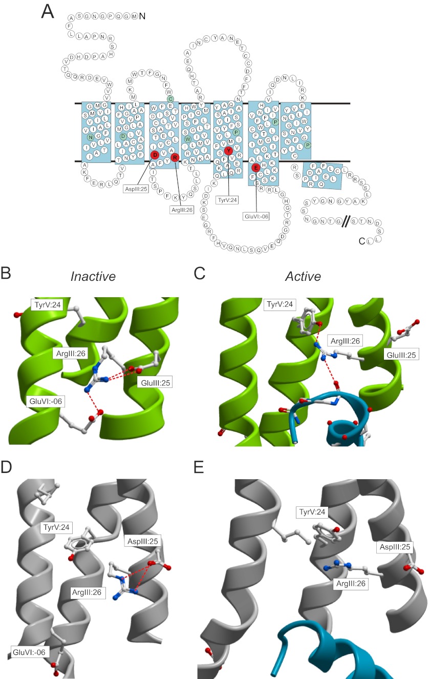
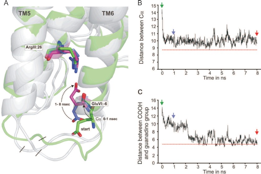
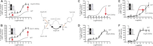
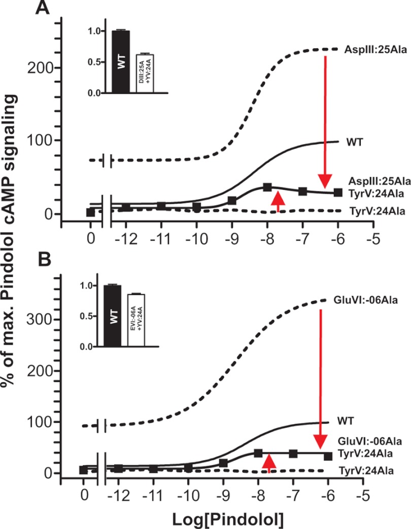
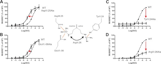
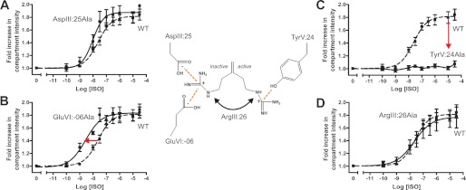
Recent high resolution x-ray structures of the β2-adrenergic receptor confirmed a close salt-bridge interaction between the suspected micro-switch residue ArgIII:26 (Arg3.50) and the neighboring AspIII:25 (Asp3.49). However, neither the expected "ionic lock" interactions between ArgIII:26 and GluVI:-06 (Glu6.30) in the inactive conformation nor the interaction with TyrV:24 (Tyr5.58) in the active conformation were observed in the x-ray structures. Here we find through molecular dynamics simulations, after removal of the stabilizing T4 lysozyme, that the expected salt bridge between ArgIII:26 and GluVI:-06 does form relatively easily in the inactive receptor conformation. Moreover, mutational analysis of GluVI:-06 in TM-VI and the neighboring AspIII:25 in TM-III demonstrated that these two residues do function as locks for the inactive receptor conformation as we observed increased G(s) signaling, arrestin mobilization, and internalization upon alanine substitutions. Conversely, TyrV:24 appears to play a role in stabilizing the active receptor conformation as loss of function of G(s) signaling, arrestin mobilization, and receptor internalization was observed upon alanine substitution of TyrV:24. The loss of function of the TyrV:24 mutant could partly be rescued by alanine substitution of either AspIII:25 or GluVI:-06 in the double mutants. Surprisingly, removal of the side chain of the ArgIII:26 micro-switch itself had no effect on G(s) signaling and internalization and only reduced arrestin mobilization slightly. It is suggested that ArgIII:26 is equally important for stabilizing the inactive and the active conformation through interaction with key residues in TM-III, -V, and -VI, but that the ArgIII:26 micro-switch residue itself apparently is not essential for the actual G protein activation.


Figures

FIGURE 1.
FIGURE 1.
The DRY motif with interaction partners in rhodopsin/opsin and the B2AR. A, serpentine model of the B2AR. The interaction partners for the ArgIII:26 micro-switch for the inactive structure, AspIII:25 and GluVI:-06, and active structure, TyrVI:24, are marked in red. B, inactive structure of rhodopsin (Protein Data Bank (PDB): 1F88) displaying the ionic lock as viewed from TM-VII. Only key residues are indicated in sticks. C, active structure of opsin (PDB: 3DQB) displaying the breakage of the ionic lock and the interaction of the TyrV:24 residue to the ArgIII:26 and again from the ArgIII.26 to the Gα-peptide. D, inactive structure of the B2AR (PDB: 2RH1). E, active structure of B2AR with Gs protein (PDB: 3SN6).
FIGURE 2.
FIGURE 2.
Molecular dynamics simulation studies of the salt bridge between ArgIII:26 of the DRY motif at the intracellular end of TM-III and GluVI:-06 at the intracellular extension of TM-VI in B2AR. A, The structure of the intracellular ends of TM-III, -V, and -VI in a molecular model of the B2AR in which T4-lysozyme was removed from the T4L-B2AR structure (2RH1) and the two ends joined. The structure shown in green is the starting structure, and structures after 1- and 8-ns simulation are also shown; in all structures, ArgIII:26, AspIII:25, TyrV:24, and GluVI:-06 are shown as sticks. Note that initially (0–1 ns), the backbone around GluVI:-06, which was in a loop-like structure, swings into a regular α-helical extension of TM-VI, and subsequently (1–8 ns), the side chain of GluVI:-06 swings over toward ArgIII:26 to form the salt bridge. B, the distance between the Cα of ArgIII:26 and GluVI:-06 as a function of simulation time. The horizontal red line indicates the distance between Cα of ArgIII:26 and GluVI:-06 in the rhodopsin structure (1GZM). C, the distance between the carbon atom in the guanidine and carboxylic acid groups of ArgIII:26 and GluVI:-06, respectively, during the molecular dynamics simulation. The horizontal red line indicates the distance between the carbon atom in the guanidine and carboxylic acid groups of ArgIII:26 and GluVI:-0g in the rhodopsin structure (1GZM).
FIGURE 3.
FIGURE 3.
Functional consequence on Gs signaling in the B2AR of Ala substitutions of the ArgIII:26/3.50 micro-switch and its potential interaction partners. A–D, basal and agonist (pindolol)-induced cAMP production in COS-7 cells transiently transfected with either WT β2-AR (dotted line) or mutant forms of the following: AspIII:25Ala (A), GluVI:-06Ala (B), TyrV:24Ala (C), and ArgIII:26Ala (D). % of max., percentage of maximum. E and F, isoproterenol-induced cAMP production in CHO-K1 cells transiently transfected with either WT (dotted line) or mutant forms of the β2-AR: TyrV:24Ala (E) and ArgIII:26Ala (F). Cell surface receptor expression, measured by enzyme-linked immunosorbent assay, is shown in the inserted column diagrams in each panel. Error bars indicate S.E.
FIGURE 4.
FIGURE 4.
Effect on Gs signaling of combining loss-of-function and gain-of-function mutants of the proposed locks for the ArgIII:26 micro-switch in B2AR. Basal and agonist (pindolol)-induced cAMP production in COS-7 cells transiently transfected with either WT (solid line) or mutant forms of B2AR is shown. A, AspIII:25Ala (dotted line), TyrV:24Ala (dotted line), and the double mutant AspIII:25Ala + TyrV:24Ala as compared with WT (solid line). % of max., percentage of maximum. B, GluVI:-06Ala (dotted line), TyrV:24Ala (dotted line), and GluVI:-06Ala+TyrV:24Ala as compared with WT (solid line). Cell surface receptor expression of the double mutants, measured by enzyme-linked immunosorbent assay, is shown in the inserted column diagrams in each panel. Error bars indicate S.D.
FIGURE 5.
FIGURE 5.
Functional consequence on β-arrestin2 mobilization measured by BRET of Ala substitution of the ArgIII:26/3.50 micro-switch and potential interaction partners. A–D, agonist (isoproterenol)-induced BRET production in COS-7 cells transiently transfected using a 1:3 ratio of either WT B2AR (dotted line)/mutant forms of the receptor and β-arrestin2. The panels show AspIII:25Ala (A), GluVI:-06Ala (B), TyrV:24Ala (C), and ArgIII:26Ala (D). Error bars indicate S.D.
FIGURE 6.
FIGURE 6.
Functional consequence on isoproterenol-induced internalization of Ala substitution of the ArgIII:26/3.50 micro-switch and potential interaction partners. A–D, agonist (isoproterenol)-induced internalization in HEK293 cells stably expressing either N-terminal SNAP-tagged WT B2AR (dotted line) or mutant forms of the receptor. The panels show AspIII:25Ala (A), GluVI:-06Ala (B), TyrV:24Ala (C), and ArgIII:26Ala (D). Error bars indicate S.D.

Similar articles

Cited by

References

    1. Jaakola V. P., Griffith M. T., Hanson M. A., Cherezov V., Chien E. Y., Lane J. R., Ijzerman A. P., Stevens R. C. (2008) The 2.6 angstrom crystal structure of a human A2A adenosine receptor bound to an antagonist. Science 322, 1211–1217 - PMC - PubMed
    1. Palczewski K., Kumasaka T., Hori T., Behnke C. A., Motoshima H., Fox B. A., Le Trong I., Teller D. C., Okada T., Stenkamp R. E., Yamamoto M., Miyano M. (2000) Crystal structure of rhodopsin: a G protein-coupled receptor. Science 289, 739–745 - PubMed
    1. Rasmussen S. G., Choi H. J., Rosenbaum D. M., Kobilka T. S., Thian F. S., Edwards P. C., Burghammer M., Ratnala V. R., Sanishvili R., Fischetti R. F., Schertler G. F., Weis W. I., Kobilka B. K. (2007) Crystal structure of the human β2 adrenergic G protein-coupled receptor. Nature 450, 383–387 - PubMed
    1. Rasmussen S. G., Choi H. J., Fung J. J., Pardon E., Casarosa P., Chae P. S., Devree B. T., Rosenbaum D. M., Thian F. S., Kobilka T. S., Schnapp A., Konetzki I., Sunahara R. K., Gellman S. H., Pautsch A., Steyaert J., Weis W. I., Kobilka B. K. (2011) Structure of a nanobody-stabilized active state of the β2 adrenoceptor. Nature 469, 175–180 - PMC - PubMed
    1. Rasmussen S. G., DeVree B. T., Zou Y., Kruse A. C., Chung K. Y., Kobilka T. S., Thian F. S., Chae P. S., Pardon E., Calinski D., Mathiesen J. M., Shah S. T., Lyons J. A., Caffrey M., Gellman S. H., Steyaert J., Skiniotis G., Weis W. I., Sunahara R. K., Kobilka B. K. (2011) Crystal structure of the β2 adrenergic receptor-Gs protein complex. Nature 477, 549–555 - PMC - PubMed

Publication types

LinkOut - more resources