. 2012 Sep 21;287(39):32922-39.
doi: 10.1074/jbc.M112.350504. Epub 2012 Jul 26.

Insights into Duffy binding-like domains through the crystal structure and function of the merozoite surface protein MSPDBL2 from Plasmodium falciparum

Affiliations

Insights into Duffy binding-like domains through the crystal structure and function of the merozoite surface protein MSPDBL2 from Plasmodium falciparum

Anthony N Hodder et al. J Biol Chem. .

Abstract

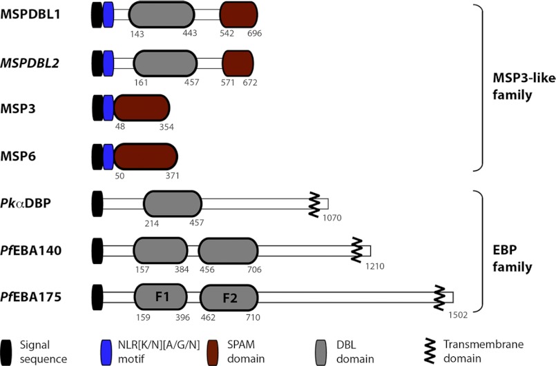
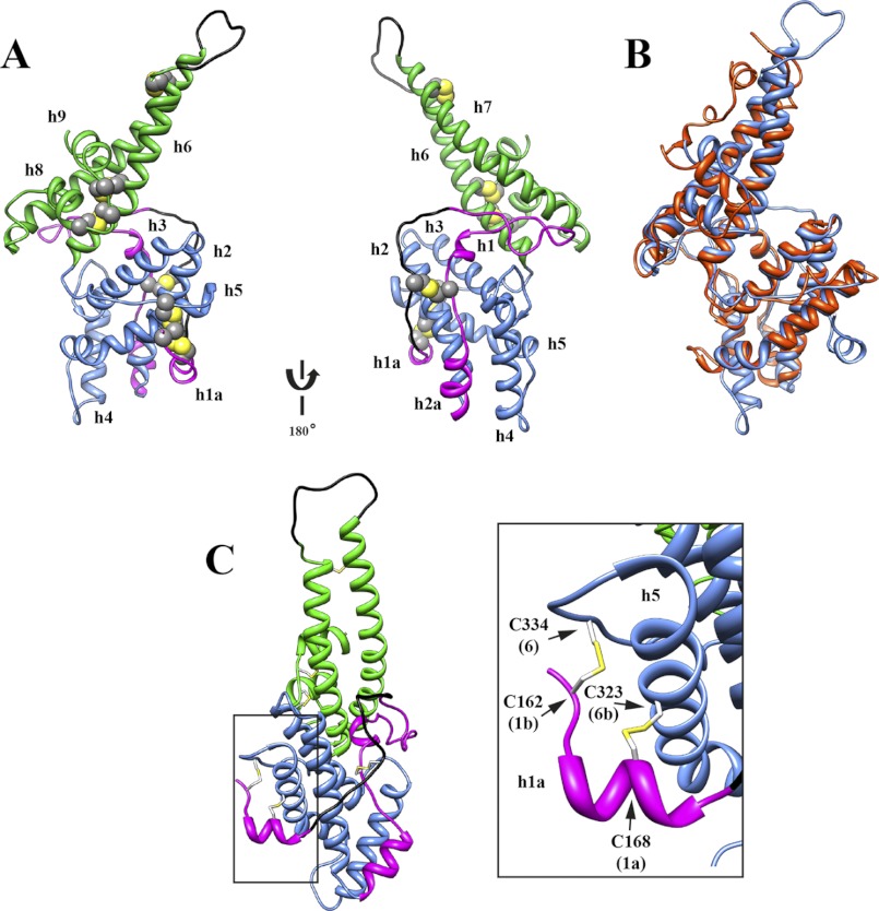
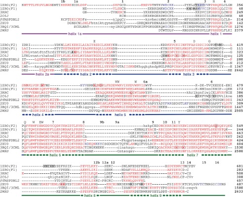
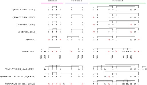
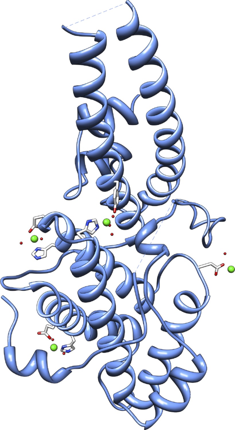
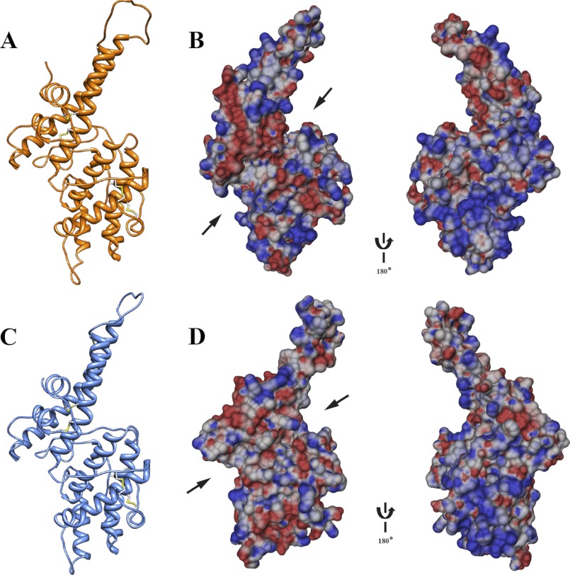
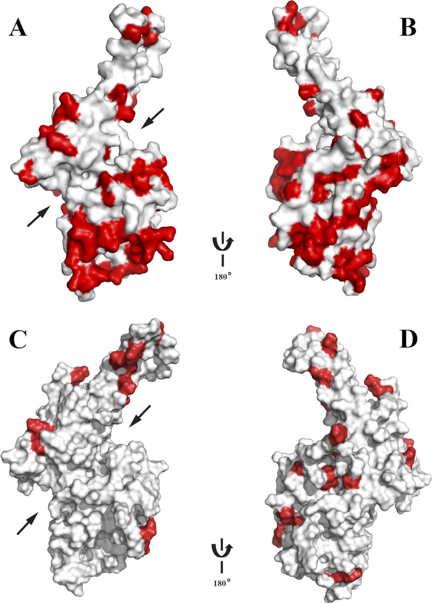
Invasion of human red blood cells by Plasmodium falciparum involves interaction of the merozoite form through proteins on the surface coat. The erythrocyte binding-like protein family functions after initial merozoite interaction by binding via the Duffy binding-like (DBL) domain to receptors on the host red blood cell. The merozoite surface proteins DBL1 and -2 (PfMSPDBL1 and PfMSPDBL2) (PF10_0348 and PF10_0355) are extrinsically associated with the merozoite, and both have a DBL domain in each protein. We expressed and refolded recombinant DBL domains for PfMSPDBL1 and -2 and show they are functional. The red cell binding characteristics of these domains were shown to be similar to full-length forms of these proteins isolated from parasite cultures. Futhermore, metal cofactors were found to enhance the binding of both the DBL domains and the parasite-derived full-length proteins to erythrocytes, which has implications for receptor binding of other DBL-containing proteins in Plasmodium spp. We solved the structure of the erythrocyte-binding DBL domain of PfMSPDBL2 to 2.09 Å resolution and modeled that of PfMSPDBL1, revealing a canonical DBL fold consisting of a boomerang shaped α-helical core formed from three subdomains. PfMSPDBL2 is highly polymorphic, and mapping of these mutations shows they are on the surface, predominantly in the first two domains. For both PfMSPDBL proteins, polymorphic variation spares the cleft separating domains 1 and 2 from domain 3, and the groove between the two major helices of domain 3 extends beyond the cleft, indicating these regions are functionally important and are likely to be associated with the binding of a receptor on the red blood cell.


Figures

FIGURE 1.
FIGURE 1.
Structure of PfMSPDBL1 and -2, MSP3, and EBL proteins. Schematic shows the important structural characteristics for PfMSPDBL1 and -2 in relation to the MSP3 and EBL protein families.
FIGURE 2.
FIGURE 2.
Expression, red cell binding, and surface localization of parasite-derived PfMSPDBL1 and -2. A, HA tagging of endogenous PfMSPDBL1 and PfMSPDBL2 proteins. Triple HA tags (HA3) were attached to the 3′ end of the PfMSPDBL1 and PfMSPDBL2 genes as described under “Experimental Procedures.” Full-length forms of the HA-tagged proteins were detected on immunoblots using a mouse anti-HA antibody. B, parasite-derived HA-tagged PfMSPDBL1 and PfMSPDBL2 proteins bind to red blood cells in an enzyme-independent manner. Post-invasion culture supernatants (Sup) from parasites expressing endogenously HA-tagged PfMSPDBL proteins or from wild type (WT) parasites, were incubated with untreated red blood cells (−) or cells treated with either trypsin (T) or neuraminidase (N) as described under “Experimental Procedures.” Bound proteins were eluted and detected on immunoblots using mouse anti-HA antibodies. Rabbit anti-EBA175 antibodies were used to confirm that the enzyme treatments had effectively removed selective red blood cell surface receptors. C, PfMSPDBL1 and -2 localize to the merozoite surface. Immunofluorescence assays of late stage schizonts and merozoites were prepared from P. falciparum (3D7 line) parasites probed with either rabbit or mouse polyclonal antibodies made to PfMSPDBL2 and marker proteins MSP119 (1st and 2nd rows), PfMSPDBL1 (3rd and 4th rows), or RON4 (5th row). 1st column, phase view; 2nd column, DAPI stained parasites; 3rd column, localization of PfMSPDBL2 (green); 4th column, localization of marker protein (red); 5th column, the merge of 3rd and 4th columns, and 6th column, the merge of columns 2–4.
FIGURE 3.
FIGURE 3.
Expression and purification of the DBL domains from PfMSPDBL2 and PfMSPDBL1. A and B show the elution profiles for PfMSPDBL1 and PfMSPDBL2, respectively, from nickel-nitrilotriacetic acid resin under denaturing conditions. C and D show SDS-PAGE analyses for the refolded DBL domains of PfMSPDBL1 and PfMSPDBL2, respectively, after ion-exchange purification. Samples were electrophoresed in sample buffer with (RD) or without (NR) reducing agent. E and F show immunoblots for PfMSPDBL1 and PfMSPDBL2 DBL domains, respectively, probed with IgG obtained from pooled hyperimmune sera from malaria endemic regions of PNG. Proteins were electrophoresed with (RD) or without (NR) reducing agent in the sample buffer prior to transfer onto PVDF membrane. G and H show reverse phase-HPLC profiles for the denatured (SM) and refolded (50) DBL domains for PfMSPDBL1 and PfMSPDBL2. See “Experimental Procedures” for additional information.
FIGURE 4.
FIGURE 4.
Red blood cell binding of recombinant DBL domains from PfMSPDBL1 and -2. A, recombinant DBL domains from PfMSPDBL1 and -2 proteins (rDBL1 and rDBL2, respectively) bind to red blood cells in an enzyme-independent manner with binding enhanced in the presence of Ca2+. Recombinant DBL domains were incubated with untreated red blood cells (−) or cells treated with either trypsin (T) or neuraminidase (N) in the presence or absence of 5 mm Ca2+, as described under “Experimental Procedures.” After passage through oil, the bound proteins were eluted with high salt, subjected to Western transfer, and then detected on immunoblots with either rabbit anti-PfMSPDBL1 or a mouse monoclonal antibody directed against the hexaHis tag of the recombinant PfMSPDBL2 DBL domain. For rDBL1 (top blot), lower bands were additionally shown (arrow) in the immunoblot as an indication of sample loading. These bands were of red blood cell protein origin and found to react only with the secondary sheep anti-rabbit IgG HRP-conjugated antibody and not the primary anti-PfMSPDBL1 antibody. Sheep anti-mouse IgG HRP antibodies used to assist in visualizing the rDBL2 (middle blot) did not cross-react with red blood cell proteins. EBA-175, obtained from parasite culture supernatants (Sup), was used to demonstrate effective trypsin and neuraminidase treatment of red blood cells. The EBA-175-binding phenotype is sensitive to treatment with either of these enzymes. An antibody targeting the C-terminal cysteine-rich region (CR) or region 6 of EBA175 was used for detection (lower blot) (51). B, binding of the recombinant DBL domains of MSPDBL1 and -2 to erythrocytes is enhanced in the presence of specific metal ions. Red blood cells were incubated with recombinant DBL domains in the presence of a selection of di- and monovalent cations (5 mm final concentration) as described under “Experimental Procedures.” Only metal ions that enhanced the binding of rDBL1 or rDBL2 to erythrocytes resulted in detectable levels of these proteins on immunoblots. Assays were performed using untreated erythrocytes and carried out as described in A and under “Experimental Procedures.” Lanes labeled rDBL1 or rDBL2 show recombinant protein not used in the binding assay. Other lanes show outcome of binding assays performed with different metal ions in conjunction with rDBL1 (upper blot) or rDBL2 (lower blot). C, binding of recombinant DBL domains of PfMSPDBL1 and -2 to red blood cells is dependent upon their disulfide bond-stabilized conformation. Red blood cells were incubated with refolded rDBL domain (rDBL1 or rDBL2) or reduced and alkylated rDBL domains (rDBL1-R/A or rDBL2-R/A) in the presence of 5 mm Ca2+ as described in A and under “Experimental Procedures.” 1st and 2nd lanes of each immunoblot contain recombinant rDBL1 or rDBL2 and rDBL1-R/A or rDBL2-R/A not used in binding assays as an identification control. 4th and 5th lanes show the recovery of the same proteins when used in binding assays. Note that the rDBL1-R/A or rDBL2-R/A proteins could not be recovered from these assays. D, effect of Ca2+ on the binding of parasite-derived, full-length PfMSPDBL2 to red blood cells can be reversed in the presence of EGTA. HA-tagged MSPDBL2 (i.e. full-length MSPDBL2-HA) derived from parasite cultures (see Fig. 2A) was used in these binding assays. 1st lane, parasite culture supernatant only, not used in the binding assay. 2nd lane, no Ca2+ added to the culture supernatant used in the binding assay. 3rd lane, 1 mm Ca2+ added to the culture supernatant and used in the binding assay. 4th lane, 10 mm EGTA in culture supernatant and binding assay. 5th lane, 1 mm Ca2+ + 1 mm EGTA in culture supernatant and binding assay. 6th lane, 1 mm Ca2+ + 2 mm EGTA in culture supernatant and binding assay. 7th lane, 1 mm Ca2+ + 5 mm EGTA in culture supernatant and binding assay. 8th lane, 1 mm Ca2+ + 10 mm EGTA in culture supernatant and binding assay; 9th lane, a wild type parasite culture supernatant control, in which MSPDBL2 does not have the C-terminal HA tag + 1 mm Ca2+ used in the binding assay. EBA-175, obtained from the same parasite culture supernatants used as a source of PfMSPDBL2, was used as a binding control under identical assay conditions. The rabbit polyclonal antibody used to detect EBA-175 targeted regions 3–5 of the molecule (R35) (51). E, histograms and dot blots obtained from FACS analyses of the binding of rDBL1 to untreated and enzyme-treated erythrocytes. Enzyme treatments and assay conditions are discussed further under “Experimental Procedures.” Error bars on histograms indicate means ± S.E. obtained from three independent assays. Recombinant PfRh4.9 was used as a binding control for enzyme-treated erythrocytes. This protein can bind to neuraminidase-treated erythrocytes but not erythrocytes treated with either trypsin or chymotrypsin. Representative dot blots are shown for each of the rDBL1 assay conditions. Numbers outside of the purple boxes refer to the percentage of erythrocytes with bound rDBL1 relative to the erythrocyte population. The various enzyme-treated erythrocytes used in the assays are indicated by the following symbols: −, no treatment; T, trypsin treatment; N, neuraminidase treatment; C, chymotrypsin treatment. F, histograms and dot blots obtained from FACS analyses of the binding of refolded and reduced and alkylated forms of rDBL1 to erythrocytes. Binding was performed on untreated erythrocytes in the presence of 1 mm Ca2+. See E and under “Experimental Procedures” for further details. A Student's t test was performed on the mean values obtained for the % binding of the refolded (rDBL1) and reduced and alkylated (rDBL-R/A) forms of rDBL1. A p < 0.001 is considered significant. G, histograms and dot blots obtained from FACS analyses of the binding of rDBL1 in the presence of increasing concentrations of EGTA. Binding was performed using untreated erythrocytes with 1 mm Ca2+ present in the binding buffer. The EGTA concentration within the binding assays ranged from 0 to 60 mm as indicated. Recombinant PfRh4.9 was used as a binding control over the EGTA concentration range used in the binding assays. See A and under “Experimental Procedures” for further details.
FIGURE 5.
FIGURE 5.
Structure of the DBL domain from PfMSPDBL2. A, ribbon diagram displaying the structure of the PfMSPDBL2 DBL domain. Disulfide bridges are highlighted in yellow. A 180° rotation of the structure is shown, and helices are labeled as depicted in Fig. 6. Regions corresponding to subdomains 1–3 are colored magenta, blue, and green, respectively. Only electron density corresponding to Cys-177 was observed in the region between residues Ser-172 and Asn-185, and no electron density was observed for the loop (Gln-375–Val-387) between helices 5 and 6. These regions were modeled using the LoopModel utility, part of the MODELLER package (31), and are colored in black. B, overlay of PfMSPDBL2-DBL (blue) with the DBL 6e domain of PfEMP1 VAR2CSA (red). C, schematic showing the disulfide linkages (yellow) between subdomains 1 (magenta) and 2 (blue) in the DBL domain of PfMSPDBL2. The depicted view is obtained by a 90° anticlockwise rotation of the first schematic in A. The PDB code for the native structure is 3VUU.
FIGURE 6.
FIGURE 6.
Structure-based sequence alignment for various DBL domains found in Plasmodium spp. Residues participating in helices are colored red, and those in β-strands are colored blue. Two cysteine residues not engaged in disulfide bond formation are underlined. Residues that were either engineered mutations or that are anomalous are presented in italics. Cysteine residues engaged in disulfide bonds and strictly conserved residues are highlighted. The location of the canonical helices is shown. Residues in lowercase for PfMSPDBL2 are not observed in the x-ray structure. Residues presented in boldface and highlighted have been implicated in the binding of substrate. Magenta, blue, and green underlines represent regions defined as subdomains 1–3, respectively. Protein identification is based on the PDB identifier code with 1ZRO = PfEBA175 DBL; 3RRC = Pv DBP DBL; 2C6J = Pk DBP DBL; 2XUO = PfEMP1-NTS-DBL1a1–VarO; 3BQI/3CML = PfEMP1 VAR2CSA DBL 3X; and 2WAU = PfEMP1 VARCSA DBL 6ϵ.
FIGURE 7.
FIGURE 7.
Comparison of the disulfide bond architectures found in various DBL domains of Plasmodium spp. Numbers 1–14 represent the relative position of cysteine residues conserved in the structure of the EBA-175 F2 DBL domain. Additional numbers and letters represent a shift in the position of cysteine residues found in other DBL domains relative to those in the EBA-175 F2 DBL domain. A red cross through numbers 1–14 represents the loss of a cysteine from a position found in EBA-175 F2 DBL domain. The identity of individual Cys residues involved in disulfide bonds within the DBL domain of PfMSPDBL2 is shown. Magenta, blue, and green lines indicate the location of Cys residues in subdomain 1–3, respectively. PDB identifier codes for each structure are shown in parentheses where known. The disulfide architecture for Pf332 is shown as described (29).
FIGURE 8.
FIGURE 8.
Structure of PfMSPDBL2 and the zinc-binding sites. The four zinc-binding sites are shown S1, His-249 and Tyr-356; S2, Glu-254 and His-257; S3, His-316 and Glu-319; S4, Asp-278, with the zinc cation represented as a green sphere. The PDB code for the structure with coordinated Zn2+ is 3VUV.
FIGURE 9.
FIGURE 9.
Modeled structures of the PfMSPDBL1 and -2 DBL domains. A, ribbon schematic of the modeled PfMSPDBL1 DBL domain. Disulfide bridges are displayed in yellow. B, electrostatic surface potential diagrams, with 180° rotation, for the DBL domain of PfMSPDBL1. C, ribbon schematic of the modeled DBL domain for PfMSPDBL2. Disulfide bridges are displayed in yellow. D, electrostatic surface potential diagrams, with 180° rotation, for the DBL domain of PfMSPDBL2. Electrostatic surface diagrams in B and D are based on the modeled structures for the DBL domains of PfMSPDBL1 and -2, including loops missing in the x-ray structure of PfMSPDBL2. Arrows indicate the location of the cleft that occurs between subdomains 2 and 3 in these DBL domains.
FIGURE 10.
FIGURE 10.
Naturally occurring polymorphisms of the PfMSPDBL2 and PfMSPDBL1 DBL domains. The structures are depicted in surface format with polymorphisms colored red. The orientation in A is as that displayed in Fig. 5A. The orientation in B is rotated 180° to that displayed in A. The orientation in C is as that displayed in Fig. 9A. The orientation in D is rotated 180° to that displayed in C. Arrows shown in A and C indicate the location of the cleft between subdomains 2 and 3 in the DBL domains of PfMSPDBL2 and -1, respectively. Coloring of the electrostatic potential is from red (−15 kJ mol−1), through white (0 kJ mol−1) to blue (+15 kJ mol−1).

Similar articles

Cited by

References

    1. Cowman A. F., Crabb B. S. (2006) Invasion of red blood cells by malaria parasites. Cell 124, 755–766 - PubMed
    1. Chitnis C. E., Miller L. H. (1994) Identification of the erythrocyte binding domains of Plasmodium vivax and Plasmodium knowlesi proteins involved in erythrocyte invasion. J. Exp. Med. 180, 497–506 - PMC - PubMed
    1. Singh S. K., Hora R., Belrhali H., Chitnis C. E., Sharma A. (2006) Structural basis for Duffy recognition by the malaria parasite Duffy-binding-like domain. Nature 439, 741–744 - PubMed
    1. Gardner M. J., Hall N., Fung E., White O., Berriman M., Hyman R. W., Carlton J. M., Pain A., Nelson K. E., Bowman S., Paulsen I. T., James K., Eisen J. A., Rutherford K., Salzberg S. L., Craig A., Kyes S., Chan M. S., Nene V., Shallom S. J., Suh B., Peterson J., Angiuoli S., Pertea M., Allen J., Selengut J., Haft D., Mather M. W., Vaidya A. B., Martin D. M., Fairlamb A. H., Fraunholz M. J., Roos D. S., Ralph S. A., McFadden G. I., Cummings L. M., Subramanian G. M., Mungall C., Venter J. C., Carucci D. J., Hoffman S. L., Newbold C., Davis R. W., Fraser C. M., Barrell B. (2002) Genome sequence of the human malaria parasite Plasmodium falciparum. Nature 419, 498–511 - PMC - PubMed
    1. Howell D. P., Levin E. A., Springer A. L., Kraemer S. M., Phippard D. J., Schief W. R., Smith J. D. (2008) Mapping a common interaction site used by Plasmodium falciparum Duffy binding-like domains to bind diverse host receptors. Mol. Microbiol. 67, 78–87 - PubMed

Publication types

MeSH terms

LinkOut - more resources