Review
. 2012 Oct 1;303(7):C699-710.
doi: 10.1152/ajpcell.00188.2012. Epub 2012 Jul 25.

Structure and activity of the acid-sensing ion channels

Affiliations
Review

Structure and activity of the acid-sensing ion channels

Thomas W Sherwood et al. Am J Physiol Cell Physiol. .

Abstract

The acid-sensing ion channels (ASICs) are a family of proton-sensing channels expressed throughout the nervous system. Their activity is linked to a variety of complex behaviors including fear, anxiety, pain, depression, learning, and memory. ASICs have also been implicated in neuronal degeneration accompanying ischemia and multiple sclerosis. As a whole, ASICs represent novel therapeutic targets for several clinically important disorders. An understanding of the correlation between ASIC structure and function will help to elucidate their mechanism of action and identify potential therapeutics that specifically target these ion channels. Despite the seemingly simple nature of proton binding, multiple studies have shown that proton-dependent gating of ASICs is quite complex, leading to activation and desensitization through distinct structural components. This review will focus on the structural aspects of ASIC gating in response to both protons and the newly discovered activators GMQ and MitTx. ASIC modulatory compounds and their action on proton-dependent gating will also be discussed. This review is dedicated to the memory of Dale Benos, who made a substantial contribution to our understanding of ASIC activity.


Figures

Fig. 1.
Fig. 1.
Overview of the acid-sensing ion channel 1 (ASIC1) crystal structure. A: surface view of trimeric chicken ASIC1 (cASIC1a) created with the UCSF Chimera package from Protein Data Bank (PDB) ID: 3HGC (98). The coloration indicates domains identified by Jasti et al. (59, 71): transmembrane domains 1 and 2 (TM1 and TM2, red); wrist (red); palm (blue); knuckle (cyan); finger (purple); thumb (green); and β-ball (orange). Notice that ASICs display a chalice shape with a large extracellular domain. The acidic pocket is located at the interface between 2 subunits and is indicated by the black box (71). B: one subunit of cASIC1 in ribbon format. Coloration indicates the specific domains identified by Jasti et al. (71): The extracellular domain also has 12 β-sheets (β1–12), 7 α-helices (α1–7), and 7 disulfide bonds (illustrated in pink). Note that one of the disulfide bonds is obscured from view by the upper region of the palm domain. The extracellular domain also contains a Cl (red sphere) binding site in the thumb domain. Images for all figures were rendered using the crystal structure of the functional cASIC1 (PDB ID: 3HGC) and the UCSF Chimera package (98). Chimera is developed by the Resource for Biocomputing, Visualization, and Informatics at the University of California, San Francisco, funded by grants from the National Institutes of Health, National Center for Research Resources (2P41RR001081) and National Institute of General Medical Sciences (9P41GM103311). [Adapted by permission from Macmillan Publishers Ltd: Nature (Ref. , copyright 2009, and Ref. , copyright 2007).]
Fig. 2.
Fig. 2.
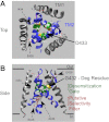
Structure of the pore region of ASIC1. The pore of cASIC1 is shown from the top (A) and side (B) view (PDB ID: 3HGC). Transmembrane domain 2 (TM2) is indicated in blue and the desensitization gate (D433-I434-G435-G436) is shown in green. Residue D433 is thought to represent the gate residue and is the most extracellular residue of the desensitization gate located in the narrowest region of the pore when the channel is desensitized (59, 86). The degenerin (Deg) residue (G432) is highlighted in gold. Mutations of this residue have long been known to alter acid-dependent gating of ASICs. A residue critical for Ca2+ block, E426 is highlighted in purple (96). The putative ion selectivity filter (G443-A444-S445) is shown in red (86). Note that only the ribbon of glycine 443 is shown in red. Images were formulated using the UCSF Chimera package (98). [Adapted by permission from Macmillan Publishers Ltd: Nature (Ref. , copyright 2009) and Nature Communications (Ref. , copyright 2011).]
Fig. 3.
Fig. 3.
The location of amino acids in the extracellular domain known to be involved in ASIC gating. On the left, subunits within the multimeric channel have domains color coded as in Fig. 1. Note that one subunit within the trimeric channel has been removed to better highlight important regions. Boxes on the right indicate higher magnification of the highlighted regions showing the locations of specific amino acid residues. A: the Cl binding site and residues of interest around the thumb domain are shown in the far right box. Note that one of the residues involved in coordinating Cl (K212) is located on the adjacent subunit which is not shown. The lower box shows Y72 and W288, critical residues in the wrist domain. The loop shown in this inset is also involved in propagating the signal from extracellular modulators [i.e., RFamides (peptide terminating in the sequence arginine-phenylalanine-amide)] to channel gating (114). Residues corresponding to those involved in GMQ/agmatine binding of ASIC3 are shown in the dashed box. These residues are located within the palm regions of the trimeric channel. Note that residue E80 from one subunit (white label) interacts with E417 from the adjacent subunit (yellow label). B: residues of interest located in the finger and knuckle domains. Note that the arrow indicates that the image in A has been rotated to the right to highlight the important region. In the crystal structure (3HGC), side chains for residues K134 and R146 were unable to be visualized. The side chains shown here were inserted using the swapaa command in Chimera software. The acidic pocket is illustrated with a dashed box. The linker region is outlined by the solid box. C: higher-magnification image of the acidic pocket which is composed of the thumb and finger of one subunit and the upper region of the palm domain from the adjacent subunit. The crystal structure in this image was rotated in relationship to the trimer shown in B to show the two carboxyl-carboxylate and one carbonyl-carboxylate pairs (D238-D350, E239-D346, and E220-D408). D: higher magnification of the linker region of one cASIC1 subunit. The crystal structure in this image was rotated in relation to the ribbon subunit shown in B to highlight the β1-β2 and β11-β12 linkers (pink) and residues of interest. Images were formulated using the UCSF Chimera package and PDB ID: 3HGC (59, 71, 98). [Adapted by permission from Macmillan Publishers Ltd: Nature (Ref. , copyright 2009, and Ref. , copyright 2007).]

References

    1. Adams CM, Price MP, Snyder PM, Welsh MJ. Tetraethylammonium block of the BNC1 channel. Biophys J 76: 1377–1383, 1999 - PMC - PubMed
    1. Adams CM, Snyder PM, Price MP, Welsh MJ. Protons activate brain Na+ channel 1 by inducing a conformational change that exposes a residue associated with neurodegeneration. J Biol Chem 273: 30204–30207, 1998 - PubMed
    1. Adams CM, Snyder PM, Welsh MJ. Paradoxical stimulation of a DEG/ENaC channel by amiloride. J Biol Chem 274: 15500–15504, 1999 - PubMed
    1. Akopian AN, Chen CC, Ding Y, Cesare P, Wood JN. A new member of the acid-sensing ion channel family. Neuroreport 11: 2217–2222, 2000 - PubMed
    1. Andrey F, Tsintsadze T, Volkova T, Lozovaya N, Krishtal O. Acid sensing ionic channels: modulation by redox reagents. Biochim Biophys Acta 1745: 1–6, 2005 - PubMed

Publication types

MeSH terms

Substances