. 2012 Oct;194(19):5305-14.
doi: 10.1128/JB.00699-12. Epub 2012 Jul 27.

The histidine kinase PdhS controls cell cycle progression of the pathogenic alphaproteobacterium Brucella abortus

Affiliations

The histidine kinase PdhS controls cell cycle progression of the pathogenic alphaproteobacterium Brucella abortus

Charles Van der Henst et al. J Bacteriol. 2012 Oct.

Abstract

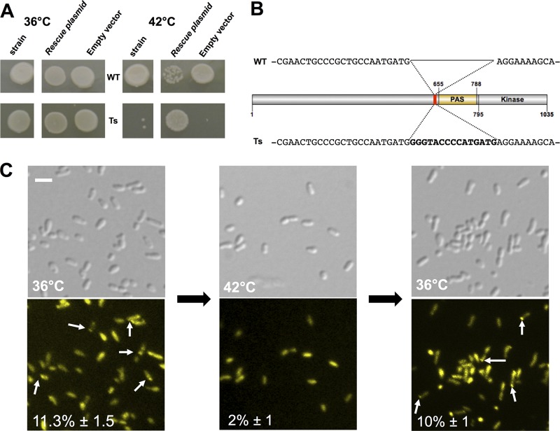
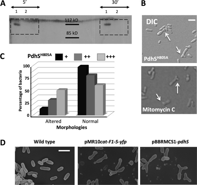
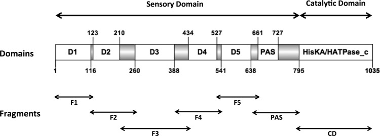
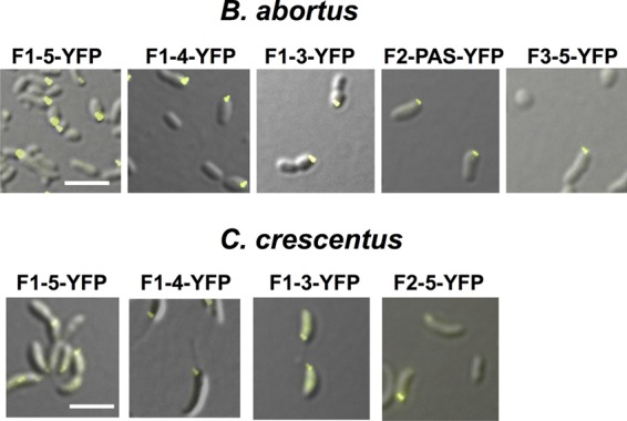
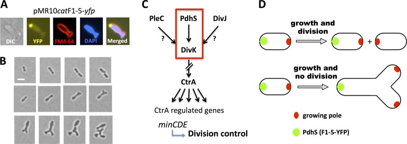
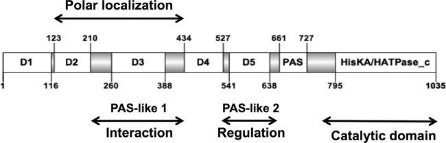
Bacterial differentiation is often associated with the asymmetric localization of regulatory proteins, such as histidine kinases. PdhS is an essential and polarly localized histidine kinase in the pathogenic alphaproteobacterium Brucella abortus. After cell division, PdhS is asymmetrically segregated between the two sibling cells, highlighting a differentiation event. However, the function(s) of PdhS in the B. abortus cell cycle remains unknown. We used an original approach, the pentapeptide scanning mutagenesis method, to generate a thermosensitive allele of pdhS. We report that a B. abortus strain carrying this pdhS allele displays growth arrest and an altered DivK-yellow fluorescent protein (YFP) polar localization at the restrictive temperature. Moreover, the production of a nonphosphorylatable PdhS protein or truncated PdhS proteins leads to dominant-negative effects by generating morphological defects consistent with the inhibition of cell division. In addition, we have used a domain mapping approach combined with yeast two-hybrid and fluorescence microscopy methods to better characterize the unusual PdhS sensory domain. We have identified a fragment of the PdhS sensory domain required for protein-protein interaction (amino acids [aa] 210 to 434), a fragment sufficient for polar localization (aa 1 to 434), and a fragment (aa 527 to 661) whose production in B. abortus correlates with the generation of cell shape alterations. The data support a model in which PdhS acts as an essential regulator of cell cycle progression in B. abortus and contribute to a better understanding of the differentiation program inherited by the two sibling cells.


Figures

Fig 1
Fig 1
The thermosensitive strain highlights a PdhS loss of function. (A) Wild-type (WT) strain of B. abortus and the thermosensitive (Ts) strain grown at the permissive temperature (36°C) or restrictive temperature (42°C), with a plasmid carrying the wild-type pdhS sequence (rescue plasmid) or the empty vector. (B) Sequence of the 15-bp scar at the insertion site of Tn4430. (C) DivK-YFP localization in the B. abortus TS strain. Differential interference contrast (DIC) images are shown in the upper panels, and YFP fluorescence is shown in the bottom panels. White arrows show polar DivK-YFP. The percentage in each panel indicates the proportion of bacteria in which a polar DivK-YFP was detected. Scale bar, 2 μm.
Fig 2
Fig 2
Several alterations of PdhS generate morphologies similar to cell division defects. (A) Autophosphorylation assay on PdhS and PdhSH805A. Recombinant PdhS-His6 (no. 1) and PdhSH805A-His6 (no. 2) proteins were used in the autophosphorylation assay, with incubations of 5 and 30 min with radioactively labeled [γ-32P]ATP as indicated. The expected size of PdhS-His6 is 111 kDa. (B) Altered morphologies generated by the overproduction of the nonphosphorylatable version of PdhS (PdhSH805A [upper panel]) and mitomycin C (lower panel), which blocks bacterial division. DIC, differential interference contrast. Scale bar, 1 μm. (C) Proportions of altered and normal morphologies when PdhSH805 is carried on an integrative plasmid (pSK; +) or on replicative plasmids, with either low (pMR; ++) or medium (pBBR; +++) copy numbers. (The number of bacteria counted for each condition was >400.) (D) Scanning electron microscopy of wild-type B. abortus (left panel), of cells producing PdhS F1-5 fused to YFP (middle panel; plasmid pMR10cat-F1-5-yfp), and cells overproducing PdhS (right panel; plasmid pBBR1MCS-pdhS). Scale bar, 2 μm.
Fig 3
Fig 3
Structural organization of PdhS. Conserved domains are shown as open boxes, and the flanking unconserved regions (UCRs) are shown as gray boxes. The numbering of the residues flanking the different regions of the protein is indicated, as well as the position of the catalytic domain (HisKA/HATPase_c) and the PAS domain. D1 to D5 designate conserved subdomains without predicted functions. The protein fragments examined in the functional assays comprise the different conserved domains with their flanking UCRs.
Fig 4
Fig 4
Interaction assay using the yeast two-hybrid (Y2H) approach. PdhS fragments that gave a positive interaction result are shown in black lines scaled to the PdhS primary structure. (A) PdhS fused to the DNA binding domain of the transcription factor Gal4 interacts with F1-CD (whole PdhS protein used as a positive control, from domain 1 to the catalytic domain), F1-3, F2-5, F2-3, F3-CD, F2-PAS, F3-4, and F3-PAS. The common fragment to all interacting fragments was F3 (residues 210 to 434). (B) FumC fused to the DNA binding domain of Gal4 interacts with the whole PdhS (F1-CD) and F1-3, F2-5, F2-3, F3-CD, F1-5, and F1-4. The common portion of the PdhS fragments interacting with FumC is F3 (residues 210 to 434). (C) PdhS fragments tested for their interaction with the response regulator DivK, reported to bind to the catalytic domain (18).
Fig 5
Fig 5
Localization assays. Shown are fragments of PdhS-YFP fusions that are able to polarly localize in B. abortus (upper panels) and in C. crescentus (lower panels). All fluorescence micrographs are merged differential interference contrast (DIC) and YFP images. Scale bar, 3 μm.
Fig 6
Fig 6
Generation of Y-shaped cells in B. abortus. (A) Differential interference contrast (DIC) and fluorescence microscopy of a representative Y-shaped cell generated by the production of F1-5-YFP. Cells were labeled with the FM4-64 and DAPI compounds (staining bacterial membranes and DNA, respectively) and observed using the fluorescent signals of YFP, FM4-64, and DAPI as indicated. (B) Time-lapse microscopy. Bacteria were placed on a slide layered with rich medium (2YT), and micrographs were taken every hour. B. abortus wild-type strain (upper panels) and B. abortus expressing the F1-5-YFP, Y-shaped cell generation (middle panels), and Y-shaped cell growth and division (lower panels). Scale bar, 1 μm. (C) Model of the regulatory pathway proposed for the interpretation of generation of altered morphologies. The red frame indicates the proposed essentiality pathway. (D) In a normal situation, without alteration of growth or division, each sibling cell inherits a new growing pole after division (shown above). If division is inhibited and growth still takes place, at a similar time or cell volume, two growing poles are also generated, but in the same cell and at the new pole, generating a Y shape (shown below).
Fig 7
Fig 7
Fragments associated with PdhS functions. PdhS fragments proposed to be required for (i) polar localization, (ii) interaction with PdhS and FumC (“Interaction”), and (iii) generation of altered morphologies (“Regulation”) are indicated. Two additional PAS-like domains are predicted, one within D3 and one within D5. The catalytic domain interacts with DivK (18).

References

    1. Bellefontaine AF, et al. 2002. Plasticity of a transcriptional regulation network among alpha-proteobacteria is supported by the identification of CtrA targets in Brucella abortus. Mol. Microbiol. 43:945–960 - PubMed
    1. Bergmiller T, Ackermann M. 2011. Pole age affects cell size and the timing of cell division in Methylobacterium extorquens AM1. J. Bacteriol. 193:5216–5221 - PMC - PubMed
    1. Brilli M, et al. 2010. The diversity and evolution of cell cycle regulation in alpha-proteobacteria: a comparative genomic analysis. BMC Syst. Biol. 4:52 doi:10.1186/1752-0509-4-52 - DOI - PMC - PubMed
    1. Brown PJ, et al. 2012. Polar growth in the Alphaproteobacterial order Rhizobiales. Proc. Natl. Acad. Sci. U. S. A. 109:1697–1701 - PMC - PubMed
    1. Brown PJ, Hardy GG, Trimble MJ, Brun YV. 2009. Complex regulatory pathways coordinate cell-cycle progression and development in Caulobacter crescentus. Adv. Microb. Physiol. 54:1–101 - PMC - PubMed

Publication types

MeSH terms

LinkOut - more resources