. 2012 Sep 1;189(5):2512-20.
doi: 10.4049/jimmunol.1102892. Epub 2012 Jul 27.

Heligmosomoides polygyrus bakeri induces tolerogenic dendritic cells that block colitis and prevent antigen-specific gut T cell responses

Affiliations

Heligmosomoides polygyrus bakeri induces tolerogenic dendritic cells that block colitis and prevent antigen-specific gut T cell responses

Arthur M Blum et al. J Immunol. .

Abstract

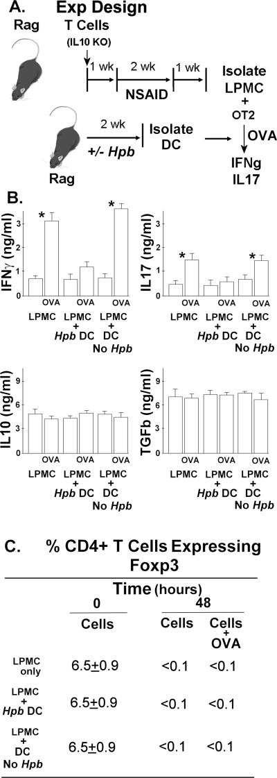
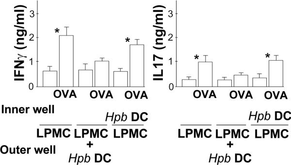
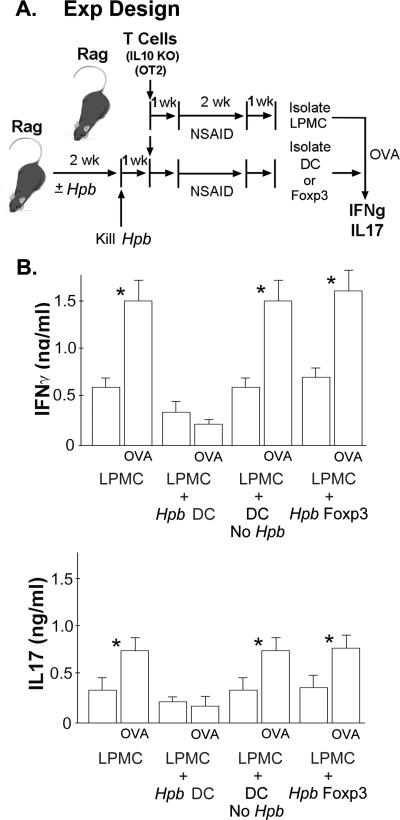
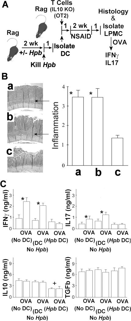
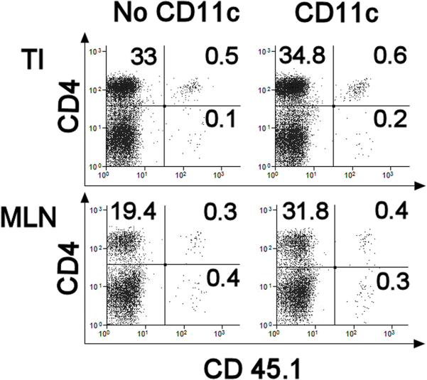
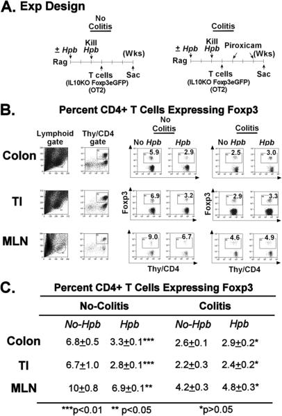
Immunological diseases such as inflammatory bowel disease (IBD) are infrequent in less developed countries, possibly because helminths provide protection by modulating host immunity. In IBD murine models, the helminth Heligmosomoides polygyrus bakeri prevents colitis. It was determined whether H. polygyrus bakeri mediated IBD protection by altering dendritic cell (DC) function. We used a Rag IBD model where animals were reconstituted with IL10⁻/⁻ T cells, making them susceptible to IBD and with OVA Ag-responsive OT2 T cells, allowing study of a gut antigenic response. Intestinal DC from H. polygyrus bakeri-infected Rag mice added to lamina propria mononuclear cells (LPMC) isolated from colitic animals blocked OVA IFN-γ/IL-17 responses in vitro through direct contact with the inflammatory LPMC. DC from uninfected Rag mice displayed no regulatory activity. Transfer of DC from H. polygyrus bakeri-infected mice into Rag mice reconstituted with IL10⁻/⁻ T cells protected animals from IBD, and LPMC from these mice lost OVA responsiveness. After DC transfer, OT2 T cells populated the intestines normally. However, the OT2 T cells were rendered Ag nonresponsive through regulatory action of LPMC non-T cells. The process of regulation appeared to be regulatory T cell independent. Thus, H. polygyrus bakeri modulates intestinal DC function, rendering them tolerogenic. This appears to be an important mechanism through which H. polygyrus bakeri suppresses colitis. IFN-γ and IL-17 are colitogenic. The capacity of these DC to block a gut Ag-specific IFN-γ/IL-17 T cell response also is significant.


Figures

Figure 1
Figure 1. Intestinal DC from Hpb-Infected Rag Mice Inhibit IFNg and IL17 Production
A. The experiment design. Rag mice were reconstituted with 106 IL10 KO T cells or 106 IL10 KO/Foxp3eGFP T cells, given i.p., and exposed to piroxicam (NSAID) to induce colitis as outlined above in panel A and in the methods section. One wk after stopping the piroxicam, Rag mice were infected with Hpb for 2 wks and then the Hpb were eliminated by treating the animals with a single orally dose of pyrantel pamoate. Age-matched control mice received the same drug treatment, but were not given the infection. FACS was used to determine the percentage of CD4+ T cells expressing Foxp3. B. Cytokines analysis. Panel B shows that adding intestinal DC from Hpb-infected mice (Hpb DC) inhibited OVA-induced cytokine secretion. Colitis LP cells cultured at 2×10⁵ cells/well in RPMI complete medium for 48h in 96-well round-bottomed plates with or without OVA (10 ug/ml) to stimulate IFNg and IL17 release. However, adding DC from mice with no prior infection (Dc No Hpb) had no such effect. Data are mean ± SE of 3-4 independent experiments For IFNg and IL17: *LPMC±OVA or LPMC + DC (No Hpb)±OVA, p<0.01. C. FACS analysis .The table in panel C shows that LPMC maintained in culture loose most of their Foxp3+ T cells and that adding Rag intestinal DC to the LPMC cultures does not maintain Foxp3+ T cell content. Data are means of 3 experiments ±SE.
Figure 2
Figure 2. DC Regulation Requires Cell Contact
The experiment was designed as outlined in figure 1, and 96 well Transwell plates were used in this experiment. Each well contained complete RPMI medium (200 ul/well). LPMC were mixed with splenic OT2 T cells (ratio 2:1) and placed in the outer chamber (2×10⁵ cells/well). In some cases, the outer chamber also contained DC (4×10⁴ cells/well) derived from TI of Rag mice infected with Hpb for 2 wks (Hpb DC). Still other wells contained Hpb DC placed in the inner chamber with the LPMC + OT2 T cells in the outer chamber separated by a 0.4 um semi-permeable membrane. Data are mean ± SE of 3 independent experiments. *LPMC±OVA; or LPMC±OVA cultured without contact with Hpb DC, p<0.01.
Figure 3
Figure 3. Hpb Infection Induces Tolerogenic DC in the Gut upon Suppression of the Colitis
A. The experiment design. To obtain LPMC from colitic mice that would respond to OVA, Rag mice first were reconstituted i.p. with 106 splenic T cells from IL10 KO mice and 3×10⁵ OT2 T cells from WT OT2 mice. Then, the Rag mice were given piroxicam (NSAID) to induce colitis. One wk after stopping the piroxicam, LPMC were isolated from the TI. To produce DC that could block the LPMC antigenic response, a second group of Rag mice were exposed to Hpb for 2 wks before reconstitution with IL10 KO T cell or IL10 KO/Foxp3eGFP reporter T cells and OT2 T cells. Then they were give piroxicam. DC or Foxp3+ T cells were isolated from the TI one wk after stopping the piroxicam. B. Cytokines analysis. Colitis LP cells cultured at 2×10⁵ cells/well in RPMI complete medium for 48h in 96-well round-bottomed plates with or without OVA (10 ug/ml) to stimulate IFNg and IL17 release. To determine if these intestinal DC or Foxp3+ T cells could modulate the LPMC cytokine response to antigen (OVA), the DC (Hpb DC) or Foxp3+ T cells (Hpb Foxp3) were added to some of the LPMC cultures at the ratio of 1:5. DC isolated from the TI of mice devoid of the infection, but otherwise manipulated similarly to the mice that received the infection, served as a source for control DC (DC No Hpb). Data are mean ± SE of 3 independent experiments. *LPMC ±OVA, LPMC+DC no Hp b ±OVA, or LPMC+Foxp3 ±OVA, p<0.01.
Figure 4
Figure 4. Adoptive Transfer of Intestinal DC from Hpb-Infected Rag Mice Protects the Recipients from Colitis
A. The experimental design. Rag mice were infected with Hpb for 2 wks and then the Hpb were eliminated by treating the animals with a single orally dose of pyrantel pamoate. Age-matched control mice received the same drug treatment, but were not given the infection. One wk after drug treatment, DC were isolated from the MLN, and the DC from either Hpb-infected or control mice were adoptively transferred by i.p. injection (4×10⁴/mouse) into Rag mice along with 4×10⁶ splenic T cells from IL10 KO mice and 2×10⁶ OT2 T cells from WT OT2 mice. A third group of Rag mice was reconstituted with IL10 KO and OT2 T cells, but received no DC. Animals then were treated with piroxicam (NSAID), as outlined above, to induce colitis. B. The histology analysis. At the end of the experiment, colonic tissue was examined microscopically to score the severity of the colitis using a 4-point scale. The data in the panel B represents the severity of inflammation in colons of mice receiving a) no DC, or were adoptively transferred b) DC from mice with no prior Hpb infection (DC No Hpb), or c) DC from mice after Hpb infection (Hpb DC). The arrows point to the intense lymphocytic infiltration in the mucosa and LP that is not present in C. Data are means ± SE from 4 separate experiments each containing 4-5 mice/group. * a or b vs. c, p<0.01. C. Cytokines analysis. Also, studied was the effect of cell transfer on mucosal cytokine production (panel C). LPMC were isolated from the TI of these very same mice. The cells were cultured in vitro at 2×10⁵ cells per well in RPMI complete medium for 48h with or without OVA (10 μg/ml) to stimulate cytokine release. Cell culture supernatants were assayed for IFNg, IL17, IL10 and TGFb using ELISAs after the 48h culture period. Data are mean ± SE of 4 independent experiments. For IFNg and IL17: *LPMC (No DC) ±OVA or LPMC (DC No Hpb ±OVA, p<0.01; LPMC (Hpb DC) ±OVA, p NS. For IL10: +Hpb DC±OVA vs. No DC or DC No Hpb, p<0.01.
Figure 5
Figure 5. DC Transfer Does not Diminish the Number of OT2 T Cells that Accumulate in the Gut or MLN
Rag mice received 4×10⁶ splenic IL10 KO T cells displaying CD45.2 and 2×10⁶ OT2 T cells expressing CD45.1 given by i.p injection. Some mice also received DC (CD11c) (4×10⁴/mouse) from the MLN of Rag mice previously infected with Hpb, while other animals did not receive DC (No CD11c). At the end of the experiment, LPMC were isolated from the TI of either the control or CD11c transfer group. Also studied were dispersed MLN cells. Cells in the lymphoid gate were subject to flow analysis to determine the relative number of LP T cells expressing CD45.1. For each group, flow analysis was performed on pooled LPMC or MLN cells isolated from 4 individual mice. Data are representative of 3 independent experiments.
Figure 6
Figure 6. Compared to WT Controls, T Cell-Reconstituted Rag Mice Infected with Hpb Do Not Display Increased Numbers of Foxp3+ CD4+ T Cells in the TI, Colon or MLN
A. The experiment design. 1) Some Rag mice were infected with Hpb (125 larvae) for 2 wks and then de-wormed by giving pyrantel pamoate. Control mice received the same drug, but no infection. Mice then received 4×10⁶ IL10KO/Foxp3 eGFP reporter T cells and 2×10⁶ OT2 T cells, by i.p. injection, one wk after pyrantel pamoate treatment. Three wks after T cell reconstitution, dispersed MLN cells as well as LPMC isolated from the colon and TI were examined using flow cytometry to assess the relative number of Foxp3+ CD4+ T cells in these cell preparations. None of these mice developed colitis, since they did not receive piroxicam (No Colitis). 2) In other experiments, mice were prepared as described above, except that the mice received 2 wks of piroxicam treatment to stimulate colitis, which only appeared in a substantial degree in the mice without prior Hpb exposure. One wk after stopping the piroxicam, the MLN cells and LPMC from the TI and colon were examined for eGFP+ Foxp3+ CD4+ T cells using flow cytometry (Colitis model). B. shows that in the no colitis model, Hpb infection actually decreased the proportion of CD4+ T cells expressing Foxp3 in the TI, colon and MLN. Compared to the no colitis group, the percentage of CD4+ T cells expressing Foxp3+ diminished following induction of colitis. Exposing mice to Hpb before induction of colitis only minimally altered the proportion of CD4+ T cells expressing Foxp3 within these tissues. Data are representative of three separate experiments. For each group, flow analysis was performed on pooled LPMC isolated from 3-4 individual mice. C. are the mean flow data from 3 individual experiments ±SE.

Similar articles

Cited by

References

    1. Weinstock JV, Summers R, Elliott DE. Helminths and harmony. Gut. 2004;53:7–9. - PMC - PubMed
    1. Elliott DE, Urban JF, Jr., Argo CK, Weinstock JV. Does the failure to acquire helminthic parasites predispose to Crohn's disease? FASEB J. 2000;14:1848–1855. - PubMed
    1. Elliott DE, Setiawan T, Metwali A, Blum A, Urban JF, Jr., Weinstock JV. Heligmosomoides polygyrus inhibits established colitis in IL-10-deficient mice. Eur. J. Immunol. 2004;34:2690–2698. - PubMed
    1. Khan WI, Blennerhasset PA, Varghese AK, Chowdhury SK, Omsted P, Deng Y, Collins SM. Intestinal nematode infection ameliorates experimental colitis in mice. Inf. Immun. 2002;70:5931–5937. - PMC - PubMed
    1. Reardon C, Sanchez A, Hogaboam CM, McKay DM. Tapeworm infection reduces epithelial ion transport abnormalities in murine dextran sulfate sodium-induced colitis. Inf. Immun. 2001;69:4417–4423. - PMC - PubMed

Publication types

MeSH terms