. 2012 Sep 1;189(5):2625-34.
doi: 10.4049/jimmunol.1102965. Epub 2012 Jul 27.

Glioma big potassium channel expression in human cancers and possible T cell epitopes for their immunotherapy

Affiliations

Glioma big potassium channel expression in human cancers and possible T cell epitopes for their immunotherapy

Lisheng Ge et al. J Immunol. .

Abstract

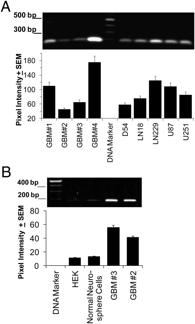
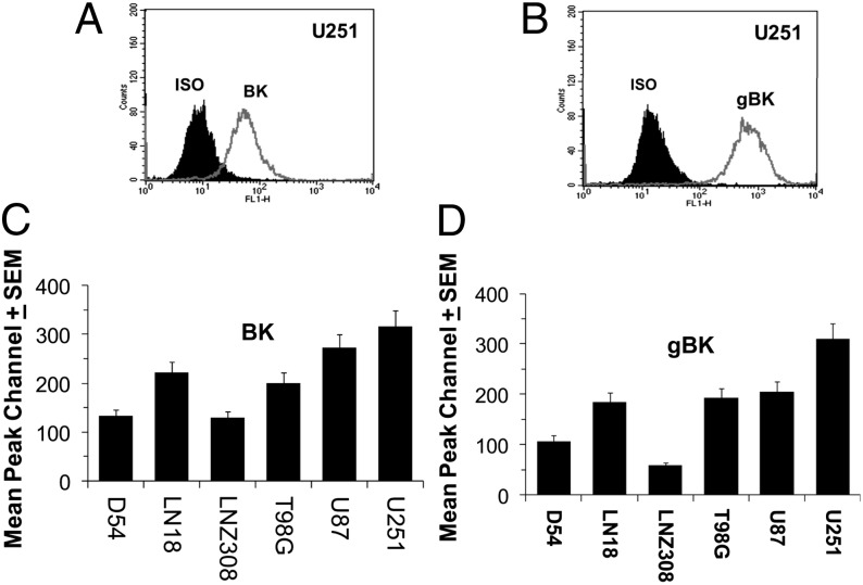
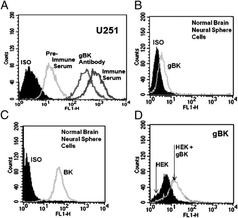
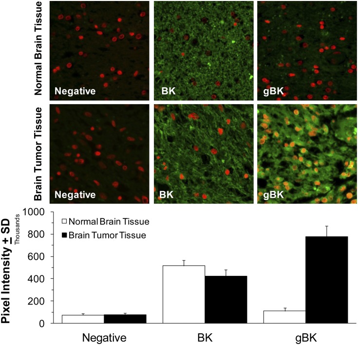
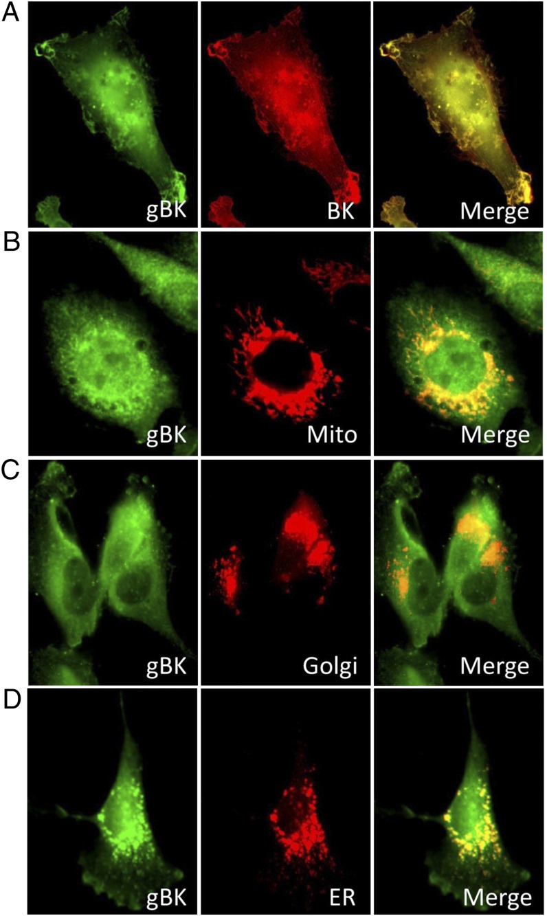
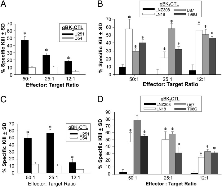
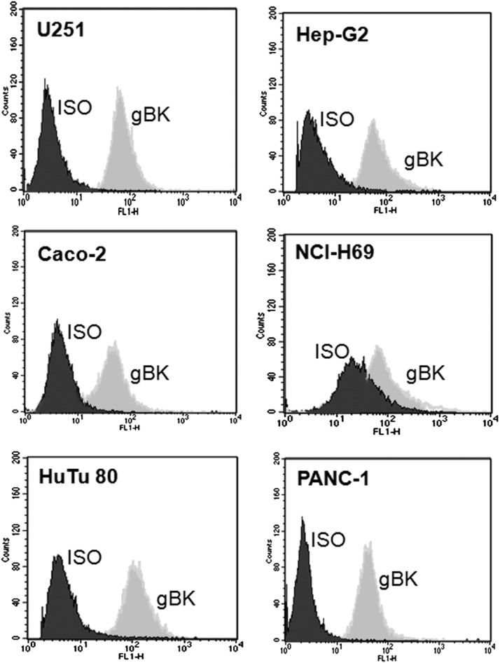
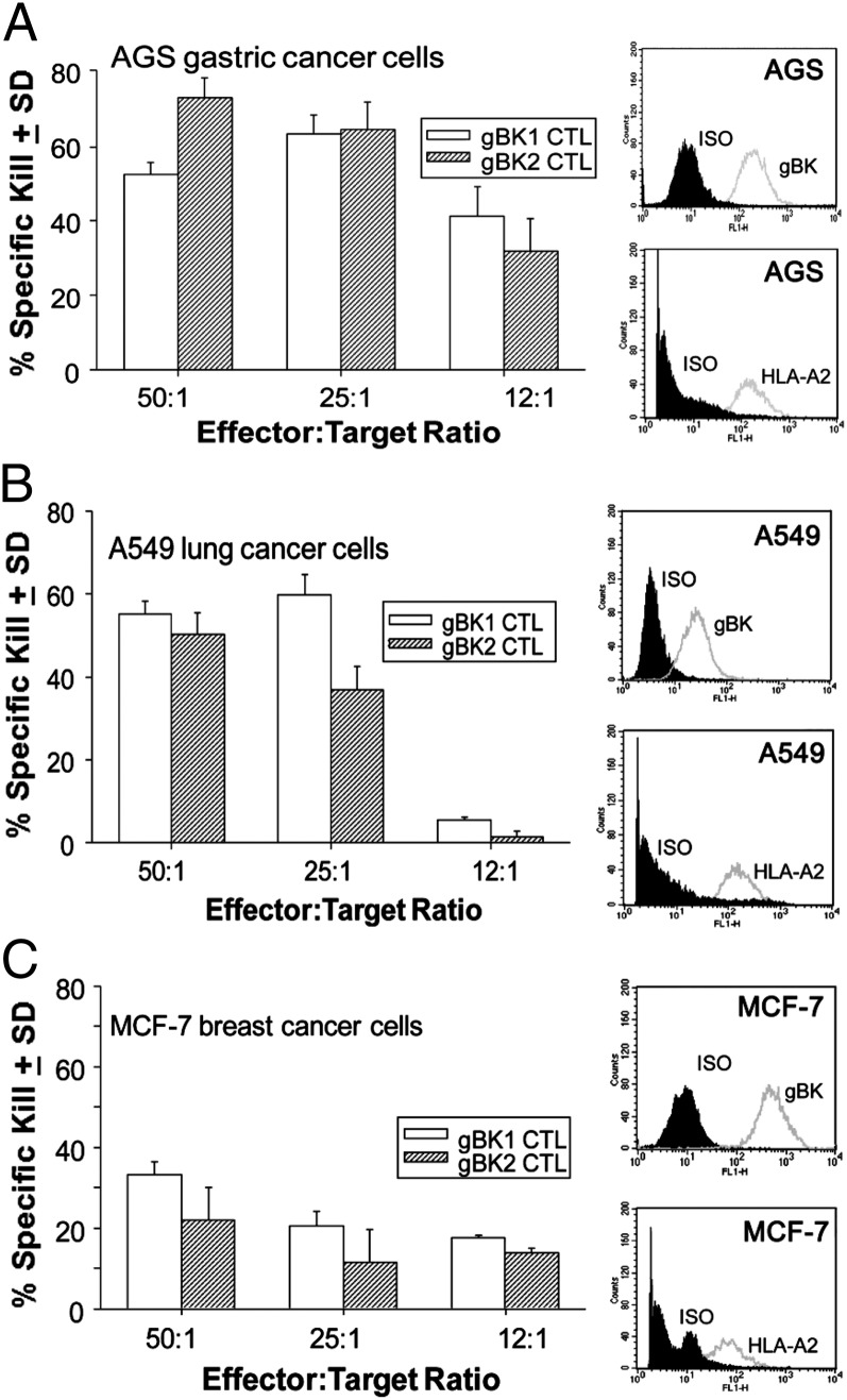
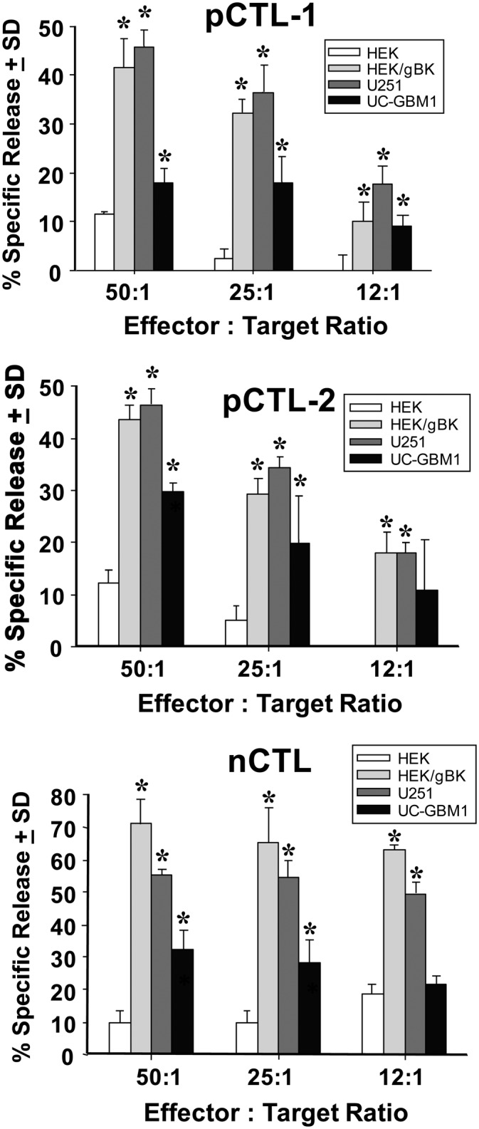
Big potassium (BK) ion channels have several spliced variants. One spliced variant initially described within human glioma cells is the glioma BK (gBK) channel. This isoform consists of 34 aa inserted into the intracellular region of the basic BK ion channel. PCR primers specific for this inserted region confirmed that human glioma cell lines and freshly resected surgical tissues from glioblastoma multiforme patients strongly expressed gBK mRNA. Normal human brain tissue very weakly expressed this transcript. An Ab specific for this gBK isoform confirmed that human glioma cells displayed this protein in the cell membrane, mitochondria, Golgi, and endoplasmic reticulum. Within the gBK region, two putative epitopes (gBK1 and gBK2) are predicted to bind to the HLA-A*0201 molecule. HLA-A*0201-restricted human CTLs were generated in vitro using gBK peptide-pulsed dendritic cells. Both gBK1 and gBK2 peptide-specific CTLs killed HLA-A2⁺/gBK⁺ gliomas, but they failed to kill non-HLA-A2-expressing but gBK⁺ target cells in cytolytic assays. T2 cells loaded with exogenous gBK peptides, but not with the influenza M1 control peptide, were only killed by their respective CTLs. The gBK-specific CTLs also killed a variety of other HLA-A*0201⁺ cancer cells that possess gBK, as well as HLA-A2⁺ HEK cells transfected with the gBK gene. Of clinical relevance, we found that T cells derived from glioblastoma multiforme patients that were sensitized to the gBK peptide could also kill target cells expressing gBK. This study shows that peptides derived from cancer-associated ion channels maybe useful targets for T cell-mediated immunotherapy.


Figures

FIGURE 1.
FIGURE 1.
RT-PCRs show the presence of gBK/hbr5 inserted sequence within clinical and human GBM cell lines. (A) RNA was isolated from four surgical specimens (GBM-1, -2, -3, and -4) and five human glioma cell lines (D54, LN18, LN229, U87, and U251). After the cDNA was synthesized, it was amplified for 40 cycles. The amplicons were loaded onto a 1.5% agarose gel and then stained with ethidium bromide and imaged (top panel). Bottom panel, Staining intensity was detected by using the UV transilluminator and then quantitated for its densitometric readings. (B) Top panel, PCR results of HEK cells, normal human neurosphere cells, and two human GBM surgical samples; bottom panel, densitometric readings.
FIGURE 2.
FIGURE 2.
Glioma cells express both BK and gBK channels as detected by intracellular flow cytometry. U251 cells were fixed, permeabilized, and stained with either the Ab toward the standard BK channel (A) or gBK-specific Ab (B). The shaded area represents the value of the isotypic controls. (C and D) Ten thousand D54, LN18, LNZ308, T98G, U87, and U251 glioma cells were analyzed by intracellular flow cytometry using either the BK or gBK Abs. The mean peak channel number and SEMs of these cell lines are presented on the y-axis. (C) BK profile. (D) gBK channel profile.
FIGURE 3.
FIGURE 3.
Specificity of the goat anti-gBK antiserum and purified anti-gBK Ab. Fixed-and-permeabilized U251 and normal human brain neurosphere cells were stained with either the preimmunized or postimmunized goat sera or with the purified goat anti-gBK Ab. Results in (A) show that (preimmunized) serum has some elevated background staining, whereas the immunized antiserum had enhanced staining. When the isotypic control and purified anti-gBK Abs were used, a lowered left shift was observed in both groups, as compared with the samples stained using whole sera. Results in (B) show the normal brain neurosphere cells to have minimal expression of gBK staining using the purified Ab, but they are positive for BK channels (C). Results in (D) show the expression of gBK by HEK cells that were either unmodified or transfected with the pcDNA/gBK plasmid for 48 h using the purified anti-gBK Ab.
FIGURE 4.
FIGURE 4.
Human GBM, but not normal brain, displays gBK. Top panel, top row, Staining profile of normal human brain; bottom row, staining profile of human GBM tissue; left column, negative control staining in the presence of a nuclear Hoescht red stain; middle column, staining with the BK channel Ab (green); right column, staining of the brain tissue using the gBK Ab (green). Original magnification ×40. Bottom panel, Summary of the average and SDs of green fluorescent staining of 40 different fields using the ImageJ intensity staining detection profile (as described in Materials and Methods).
FIGURE 5.
FIGURE 5.
gBK channels are found in the same cellular locations as the BK channels. U251 glioma cells were attached to glass cover slips overnight and fixed and stained with the anti-gBK channel Ab. Left column, gBK (green) staining of different U251 cells. (A) Middle panel, staining profile with a BK channel Ab (red); right panel, merged image. Yellow indicates that the proteins are located very close (100 nm) to each other. (B) Staining patterns when the cells were stained with the Mito-Tracker. (C) Golgi staining profile (Golgi tracker). (D) Cells stained with calreticulin, which specifically stained the ER (red). Original magnification ×100.
FIGURE 6.
FIGURE 6.
gBK-specific CTLs will kill HLA-0201+ gliomas that also express gBK. gBK1- and gBK2-specific T cells were generated and tested against either LN18, T98G, U87, or U251 glioma (HLA-A0201+/gBK+) or D54 cells (HLA-A1+/A3+, gBK+) and LNZ308 (HLA-A24+, gBK+) in a 6-h assay. (A and B) Results of one representative study done three times that shows the results of the gBK1-specific CTLs. (C and D) Results generated with the gBK2-specific CTLs. *p < 0.05 from the non-HLA-A2 control values.
FIGURE 7.
FIGURE 7.
T2 cells loaded with exogenous gBK peptides are specifically killed by the gBK1- or gBK2-specific CTLs. gBK1- or gBK2-specific CTLs were generated in vitro and then tested against T2 cells that were separately loaded with three different exogenous peptides (1 μM) for 1 h at 37°C. The cells were washed and then labeled with [51Cr] for another hour at 37°C. The cells were then washed three times and used as target cells. (A) Data generated when gBK1-specific CTLs were used. (B) Data when gBK2-specific CTLs were tested against the various T2 cells. Representative data from three different experiments are shown. *p < 0.05 from the influenza M1 control peptide (FLU) cytotoxicity values.
FIGURE 8.
FIGURE 8.
gBK can be found on a variety of other types of human tumor cell lines. Caco-2 (colon cancer), HuTu80 (duodenal adenocarcinoma), HepG2 (hepatocellular carcinoma), NCI-H69 (small cell lung cancer) and Panc-1 (pancreatic cancer) cells were fixed, permeablized, and stained with the anti-gBK Ab. Afterward, the cells were washed and stained with the anti-goat FITC labeled Ab. Ten thousand stained cells were then analyzed. The U251 cells were supplied as a reference point for staining intensity.
FIGURE 9.
FIGURE 9.
gBK-specific CTLs will also kill AGS gastric cancer, A549 lung cancer and MCF-7 breast cancer cells. gBK1 and gBK2 CTLs were tested against these HLA-A2+/gBK+ cancer cells in a 6-h [51Cr]-release assay. Right column, Intracellular flow cytometry results using the gBK Ab and the cell-surface HLA-A2 flow cytometric profile showing the intensity of this Ag on nonpermeabilized cancer cells. The shaded area represents the value of the isotypic controls. Left column, Effects of gBK1- and gBK2-specific CTLs in 6-h assays. The cytolytic activities against the AGS (A) and MCF-7 (C) cells were performed on the same day, whereas the assay with the A549 adenocarcinoma cells (B) was done 1 wk after being stimulated with immobilized anti-CD3 and anti-CD28 beads.
FIGURE 10.
FIGURE 10.
Cytotoxicity experiments with gBK-specific CTL effectors and gBK-expressing and nonexpressing target cells. pCTL-1 and pCTL-2 were generated from GBM patients and nCTLs were stimulated from a normal donor. All CTLs originated from HLA-A0201+ individuals. The DCs isolated from the PBMCs were pulsed with the gBK2 peptide prior to in vitro DC maturation. Autologous T cells were then stimulated and further expanded. The target cells included the U251 glioma cell line, early passage (P2) UC-GBM1 glioma cells (autologous to the pCTL-1), and HLA-0201+ HEK cells that were either nontransfected or transfected with the gBK plasmid 48 h prior to the assay. The gBK profile of the HEK cells is shown in Fig. 3D. All test symbols represent data that were done in quadruplicate cultures. Top panel, Results using the pCTL-1; middle panel, data generated with the pCTL-2; bottom panel, results using the nCTLs. *p < 0.05 from the nontransfected HEK control values.

References

    1. Rao R. V., Ellerby H. M., Bredesen D. E. 2004. Coupling endoplasmic reticulum stress to the cells death program. Cell Death Differ. 11: 372–380 - PubMed
    1. Jambrina E., Alonso R., Alcalde M., del Carmen Rodríguez M., Serrano A., Martínez-A C., García-Sancho J., Izquierdo M. 2003. Calcium influx through receptor-operated channel induces mitochondria-triggered paraptotic cell death. J. Biol. Chem. 278: 14134–14145 - PubMed
    1. Cassel D., Katz M., Rotman M. 1986. Depletion of cellular ATP inhibits Na+/H+ antiport in cultured human cells: modulation of the regulatory effect of intracellular protons on the antiporter activity. J. Biol. Chem. 261: 5460–5466 - PubMed
    1. Schneider D., Gerhardt E., Bock J., Müller M. M., Wolburg H., Lang F., Schulz J. B. 2004. Intracellular acidification by inhibition of the Na+/H+-exchanger leads to caspase-independent death of cerebellar granule neurons resembling paraptosis. Cell Death Differ. 11: 760–770 - PubMed
    1. Sperandio S., de Belle I., Bredesen D. E. 2000. An alternative, nonapoptotic form of programmed cell death. Proc. Natl. Acad. Sci. USA 97: 14376–14381 - PMC - PubMed

Publication types

MeSH terms

Substances