

An unusual cause of biliary obstruction
- PMID: 22844197
- PMCID: PMC3399432
- DOI: 10.4137/CCRep.S9875
An unusual cause of biliary obstruction
Abstract
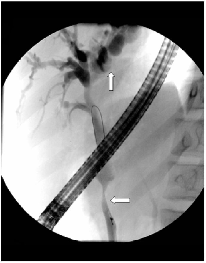
Portal biliary ductopathy (PBD) is a condition in which biliary and pancreatic ducts are extrinsically compressed by collateral branches of the portal venous system, which in turn have become dilated and varicosed due to portal hypertension. While the majority of patients with PBD are asymptomatic, a minority can present with symptoms of biliary obstruction and cholangitis with the potential of developing secondary chronic liver disease. This paper reports the case of a 29 year old male presenting with acute cholangitis, in whom PBD was diagnosed radiologically. A brief review of current literature regarding the diagnosis and management of this condition will also be presented.
Keywords: MRCP; biliary obstruction; biliary stenting Yeoh; cholangitis; portal hypertension.
Figures


Similar articles
-
How can portal vein cavernous transformation cause chronic incomplete biliary obstruction?World J Gastroenterol. 2012 Jul 14;18(26):3375-8. doi: 10.3748/wjg.v18.i26.3375. World J Gastroenterol. 2012. PMID: 22807606 Free PMC article. Review.
-
Portal Hypertensive Biliopathy: An Infrequent Cause of Biliary Obstruction.GE Port J Gastroenterol. 2015 Mar 18;22(2):65-69. doi: 10.1016/j.jpge.2015.01.003. eCollection 2015 Mar-Apr. GE Port J Gastroenterol. 2015. PMID: 28868376 Free PMC article.
-
Liver transplantation for "mass-forming" sclerosing cholangitis after laparoscopic cholecystectomy.Int J Surg Case Rep. 2013;4(10):907-10. doi: 10.1016/j.ijscr.2013.07.021. Epub 2013 Aug 3. Int J Surg Case Rep. 2013. PMID: 23995476 Free PMC article.
-
Portal cavernoma cholangiopathy: consensus statement of a working party of the Indian national association for study of the liver.J Clin Exp Hepatol. 2014 Feb;4(Suppl 1):S2-S14. doi: 10.1016/j.jceh.2014.02.003. Epub 2014 Feb 25. J Clin Exp Hepatol. 2014. PMID: 25755591 Free PMC article. Review.
-
Pseudosclerosing cholangitis in extrahepatic portal venous obstruction.Gut. 1992 Feb;33(2):272-6. doi: 10.1136/gut.33.2.272. Gut. 1992. PMID: 1541425 Free PMC article.
References
-
- Hunt AH. Compression of the Common Bile-Duct by an Enlarging Collateral Vein in a Case of Portal Hypertension. Br J Surg. 1965 Aug;52:636–7. - PubMed
-
- Bechtelsheimer H, Conrad A. Morphology of cavernous transformation of the portal vein (author’s transl) Leber Magen Darm. 1980 Apr;10(2):99–106. - PubMed
Publication types
LinkOut - more resources
Full Text Sources
