. 2012;8(7):e1002833.
doi: 10.1371/journal.pgen.1002833. Epub 2012 Jul 26.

Drosophila fatty acid transport protein regulates rhodopsin-1 metabolism and is required for photoreceptor neuron survival

Affiliations

Drosophila fatty acid transport protein regulates rhodopsin-1 metabolism and is required for photoreceptor neuron survival

Pierre Dourlen et al. PLoS Genet. 2012.

Abstract

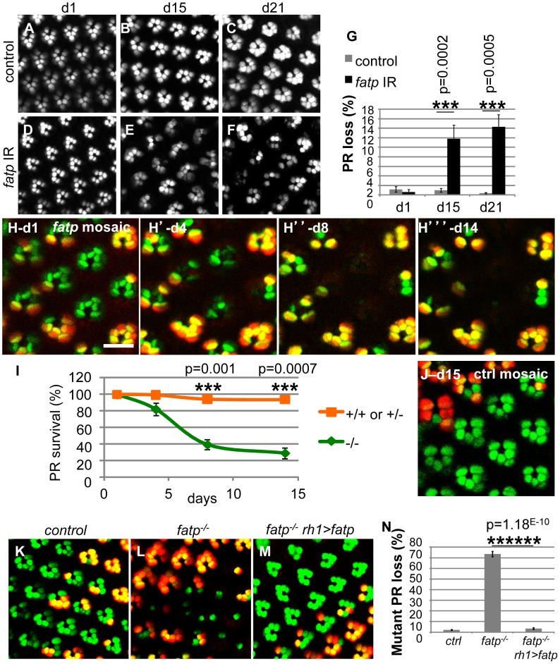
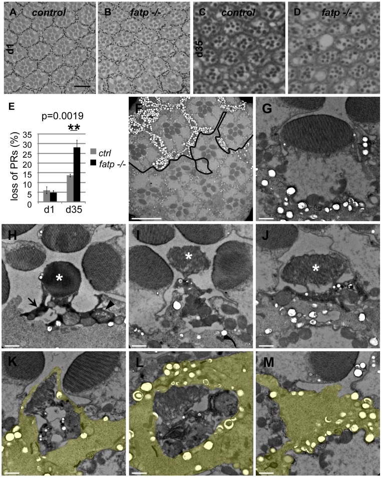
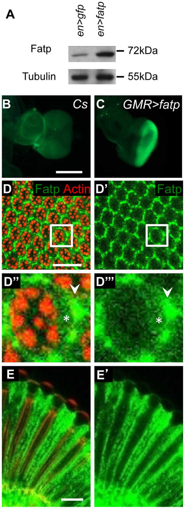
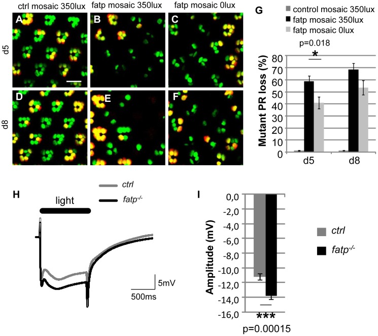
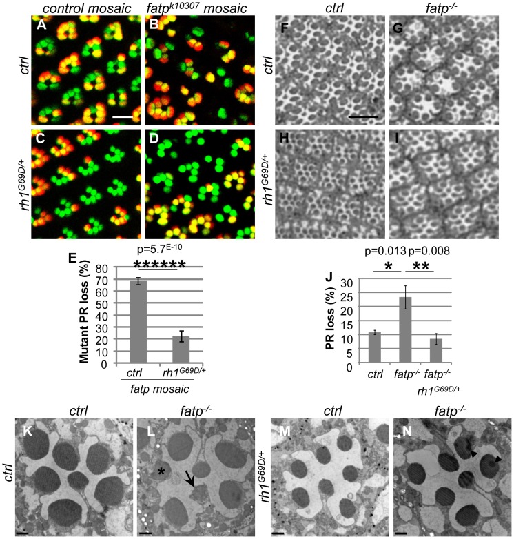
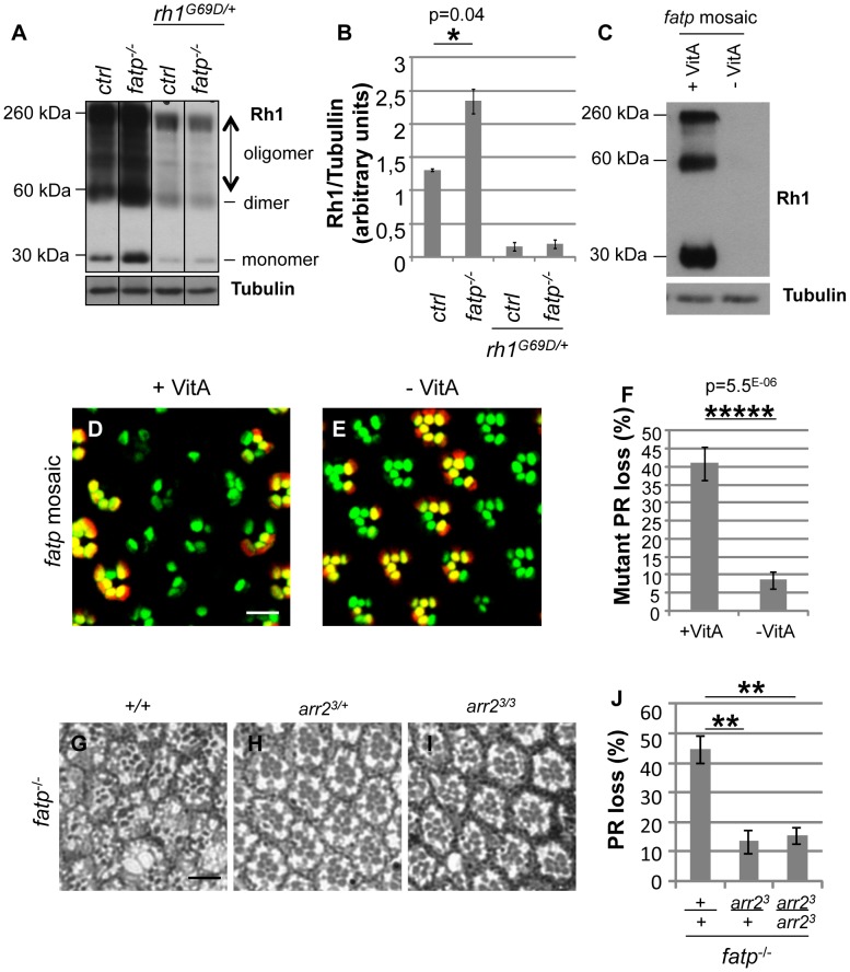
Tight regulation of the visual response is essential for photoreceptor function and survival. Visual response dysregulation often leads to photoreceptor cell degeneration, but the causes of such cell death are not well understood. In this study, we investigated a fatty acid transport protein (fatp) null mutation that caused adult-onset and progressive photoreceptor cell death. Consistent with fatp having a role in the retina, we showed that fatp is expressed in adult photoreceptors and accessory cells and that its re-expression in photoreceptors rescued photoreceptor viability in fatp mutants. The visual response in young fatp-mutant flies was abnormal with elevated electroretinogram amplitudes associated with high levels of Rhodopsin-1 (Rh1). Reducing Rh1 levels in rh1 mutants or depriving flies of vitamin A rescued photoreceptor cell death in fatp mutant flies. Our results indicate that fatp promotes photoreceptor survival by regulating Rh1 abundance.


Conflict of interest statement

The authors have declared that no competing interests exist.

Figures

Figure 1
Figure 1. Loss of fatp induces adult-onset progressive degeneration of PRs.
(A–G) Consequences of fatp knockdown by RNA interference on PR viability. (A, B, C) Visualization of PRs using a cornea-neutralization method for 1-, 15- and 21-day-old PRs expressing lacZ as a control. (D, E, F) PRs expressing the fatp-interfering RNA and (G) PR quantification. Whereas all PRs were present in control retina and fatp-knocked-down retina at day one (A, D), there were significant PR losses at days 15 and 21 in fatp-knocked-down retina (B vs E, C vs F, t-test, n≥6). (H) Time-course analysis of fatpk10307 mutant mosaic retina using the Tomato/GFP-FLP/FRT method and (I) quantification. All outer PRs (R1–R6) expressed GFP (green). FLP-mediated mitotic mutant clones were visualized by the absence of rh1-tdTomato (red). Clones of the same eye were visualized in the same fly at day one, four, eight and fourteen after hatching. (H) All PRs were present at day one (scale bar = 10 µm). (H′) From day four, mutant PRs (in green) started to disappear. (H″ and H′″) Losses of mutant PRs were significant at day eight and day fourteen (paired t-test, n = 3). (I) Quantified results are expressed as mean ± SD. (J) Analysis of a control 15-day-old mosaic retina using the Tomato/GFP-FLP/FRT method. All PRs are present. (K–N) Rescue of fatpk10307-mutant PRs by re-expression of wild-type fatp. (K) Tomato/GFP-FLP/FRT visualization of 10- to 14-day-old control, (L) fatpk10307/k10307 and (M) fatpk10307/k10307+rh1>fatp mosaic retinas and (N) quantification (t-test, n> = 5). In the control (K), all PRs were present. In fatp mutant mosaic retina (L), 70% of the mutant PRs were lost. In these retinas, mutant PRs were rescued by re-expression of fatp under the control of the rh1 promoter (M).
Figure 2
Figure 2. Histological analysis of fatp−/− PR degeneration.
(A–E) Analysis of fatpk10307 mutant PR survival using resin-embedded tangential sections. (A, C) Control retina of 1- and 35-day-old flies (scale bar = 10 µm). (B, D) Homozygous fatpk10307 retina of 1-day-old and 35-day-old flies. (E) PR losses in fatp mutant retinas were significant at 35 days compared to those in control retinas (t-test, n = 6). (F–M) Electron microscopy analysis of fatp−/− PR degeneration. fatpk10307 mosaic retina of 15-day-old flies were analyzed. (F) Wild-type and heterozygous clones were marked with numerous large pigment granules in the IOCs and at the basis of PR rhabdomeres (white spots, above the black line) whereas homozygous mutant part exhibited rare small pigment granules (under the black line, scale bar = 10 µm). Several PRs were missing in the homozygous mutant part. (G–M) Different stages of fatpk10307/k10307 PR degeneration (scale bar = 1 µm). (G) fatpk10307/k10307 PR exhibiting no sign of degeneration. (H) The cytoplasm of fatpk10307/k10307 PRs shrank and became electron-dense (arrow). Some mitochondria were swelling (arrowhead). The rhabdomere was not much affected (*). (I–J) PRs then disintegrated. This was clearly visible at the level of the rhabdomere (*). (K, L, M) Degenerating PRs were finally phagocytosed and digested by the neighboring interommatidial cell (yellow).
Figure 3
Figure 3. fatp is expressed in the adult retina.
(A) Western blot analysis of en-GAL4 UAS-GFP (en>GFP) and en-GAL4 UAS-fatp (en>fatp) adult eye extracts using anti-Fatp C11-7 and anti-tubulin antibodies. Anti-Fatp C11-7 detects a single band at 72 kDa, which corresponds to the predicted molecular weight of Fatp. Endogenous and ectopically expressed Fatp were detected. Tubulin was used as a loading control. (B, C) Immunofluorescent detection of Fatp in wild-type (CS) and GMR-GAL4 UAS-fatp (GMR>fatp) third instar larva eye imaginal disc using the anti-Fatp C11-7 antibody (scale bar = 200 µm). (C) Fatp was detected in the posterior part of the disc, which corresponds to the expression profile of the GMR promoter. (D–E) Immunostaining of endogenous Fatp and Actin in whole-mount cnbw adult retina using the anti-Fatp C11-7 antibody and phalloidin. In the tangential (D) and longitudinal plans (E), Fatp immunostaining is detected in the cytoplasm of PRs (star) and accessory cells (arrowhead). Rhabdomeres are stained with phalloidin (scale bar = 20 µm).
Figure 4
Figure 4. The visual response is altered in fatp mutant PRs.
(A–F) Tomato/GFP-FLP/FRT visualization of fatpk10307 mutant mosaic retinas from 5- and 8-day-old flies reared from the pupal stage in normal room light (350 lux) or in darkness (0 lux) (scale bar = 10 µm). (G) Quantification of fatpk10307 mutant PR loss. (A, D) No PR losses occurred in control mosaic retinas from 5-day-old and 8-day-old flies reared in room light conditions. (B, E, G) In fatp mutant retinas from light-reared flies, 58.8±4.5% and 68.5±4.9% of the mutant PRs were lost in 5-day-old and 8-day-old flies, respectively. (C, F, G) PR losses were significantly lower in flies reared in the dark (t-test, n = 8). (H) ERG recordings from control and fatpk10307/k10307 8-day-old retinas and (I) quantification. Retinas were exposed to a one second flash of orange light. (I) The amplitude of the plateau is significantly higher in the fatp mutant retina than in the control retina (t-test, n = 24).
Figure 5
Figure 5. Rh1G69D rescues PR viability in the fatp mutant.
(A–E) Analysis of the survival of control, fatpk10307, rh1G69D/+ and fatpk10307rh1G69D/+ double mutant PRs using the Tomato/GFP-FLP/FRT method in 15-day-old flies (scale bar = 10 µm). (E) Quantification of mutant PR losses (t-test, n≥12). PR loss in the fatpk10307 rh1G69D/+ double mutant was dramatically lower than in the fatp single mutant. (F–J) Analysis of the survival of control, fatpk10307, rh1G69D/+ and fatpk10307 rh1G69D/+ double mutant PRs using resin-embedded tangential sections in 28-day-old flies (scale bar = 10 µm). (J) Quantification of PR loss. Significantly more PRs died in the fatp mutant than in control retina (t-test, n = 6). In the double mutant, PR losses were reduced to control levels. (K–N) Electron microscopy analysis of whole-eye control (K), fatpk10307 (L), rh1G69D/+ (M) and fatpk10307 rh1G69D/+ (N) mutants in 28-day-old flies (scale bar = 1 µm). Whereas PRs degenerated in fatpk10307 ommatidia (arrow) and were phagocytosed by IOCs (*), PRs survived in fatpk10307 rh1G69D/+ double mutant flies. Rhabdomeres of rh1G69D/+ and fatpk10307 rh1G69D/+ outer PRs were reduced in size. Arrowheads show artifactual shadows on the sample.
Figure 6
Figure 6. Elevated Rh1 levels are responsible for PR loss in the fatp mutant.
(A) Western blot analysis of Rh1 in boiled head extracts from control, fatpk10307/k10307, rh1G69D/+ and fatpk10307/k10307/rh1G69D/+ 1 to 11-day-old flies. Tubulin was used as a loading control. (B) Quantification of protein levels. Dimer and oligomer forms of Rh1 were due to the boiling of the extracts. Rh1 levels were twofold higher in the fatp mutant than in the control. The level of Rh1 was substantially lower in the rh1G69D/+ single mutant and in the fatpk10307/k10307/rh1G69D/+ double mutant. (C) Western blot analysis of Rh1 and tubulin in fatp mutant mosaic retina extracts from 5-day-old flies reared from the embryonic stage on control (+VitA) and vitamin A-deficient (-VitA) media. Tubulin was used as a loading control. Rh1 was not detectable in flies reared on vitamin A-deficient medium. (D, E) Visualization of fatp mutant mosaic retina of 5-day-old flies reared from the embryonic stage on control (D) and vitamin A-deficient (E) media using the Tomato/GFP-FLP/FRT method (scale bar = 10 µm). (F) Quantification of mutant PR loss (t-test, n≥12). fatp mutant PR viability was dramatically restored in flies reared on vitamin A-deficient medium. (G–J) Analysis of the loss of fatpk10307, fatpk10307 arr23/+ and fatpk10307 arr23/3 double mutant PRs using resin-embedded tangential sections in 34-day-old flies (scale bar = 10 µm). (H) Quantification of PR loss. In the double mutants retinas, PRs were significantly rescued (p = 0.0021 and p = 0.0016, t-test, n = 6).

Similar articles

Cited by

References

    1. Sohocki MM, Daiger SP, Bowne SJ, Rodriquez JA, Northrup H, et al. Prevalence of mutations causing retinitis pigmentosa and other inherited retinopathies. Hum Mutat. 2001;17:42–51. - PMC - PubMed
    1. Lee ES, Flannery JG. Transport of truncated rhodopsin and its effects on rod function and degeneration. Invest Ophthalmol Vis Sci. 2007;48:2868–2876. - PMC - PubMed
    1. Mollereau B, Domingos PM. Photoreceptor differentiation in Drosophila: from immature neurons to functional photoreceptors. Dev Dyn. 2005;232:585–592. - PubMed
    1. Wang T, Montell C. Phototransduction and retinal degeneration in Drosophila. Pflugers Arch. 2007;454:821–847. - PubMed
    1. Shieh BH. Molecular genetics of retinal degeneration: A Drosophila perspective. Fly (Austin) 2011;5 - PMC - PubMed

Publication types

MeSH terms