. 2012 Apr;77(2):22-34.
doi: 10.12659/pjr.882967.

Evaluation of solitary pulmonary nodule detected during computed tomography examination

Affiliations

Evaluation of solitary pulmonary nodule detected during computed tomography examination

Agnieszka Choromańska et al. Pol J Radiol. 2012 Apr.

Abstract

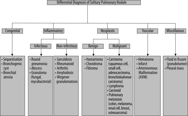
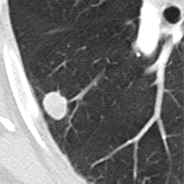
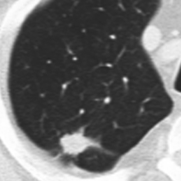
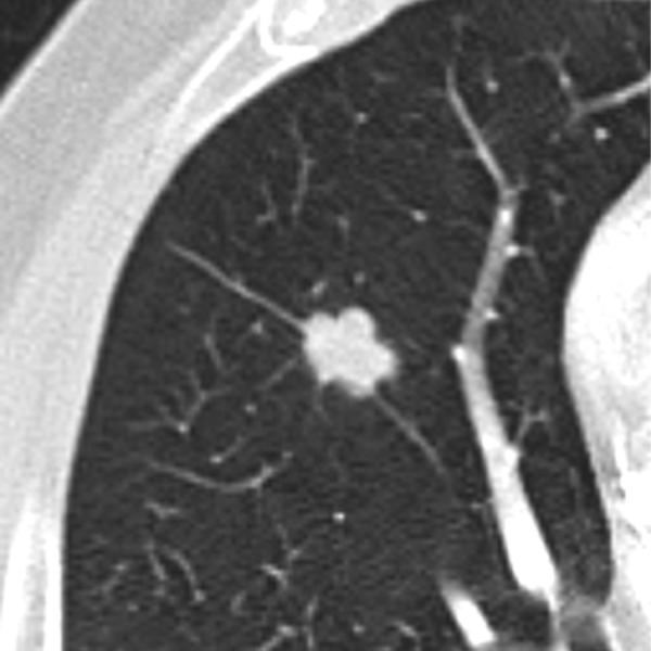
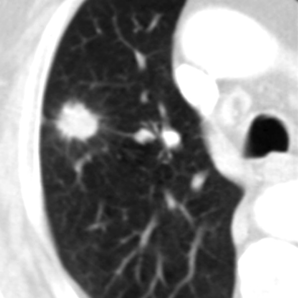
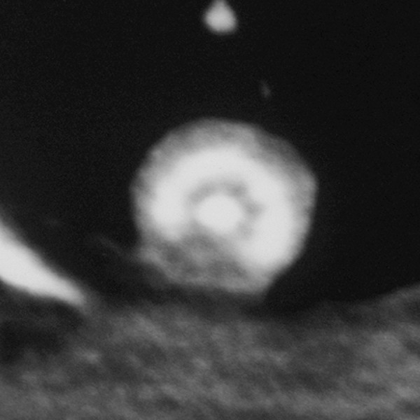
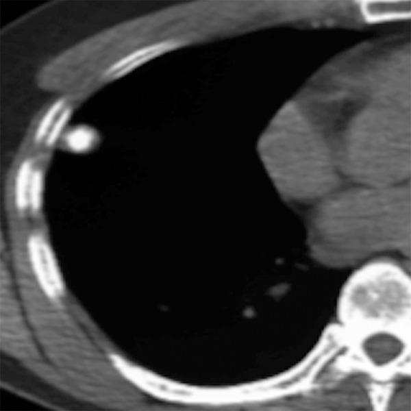
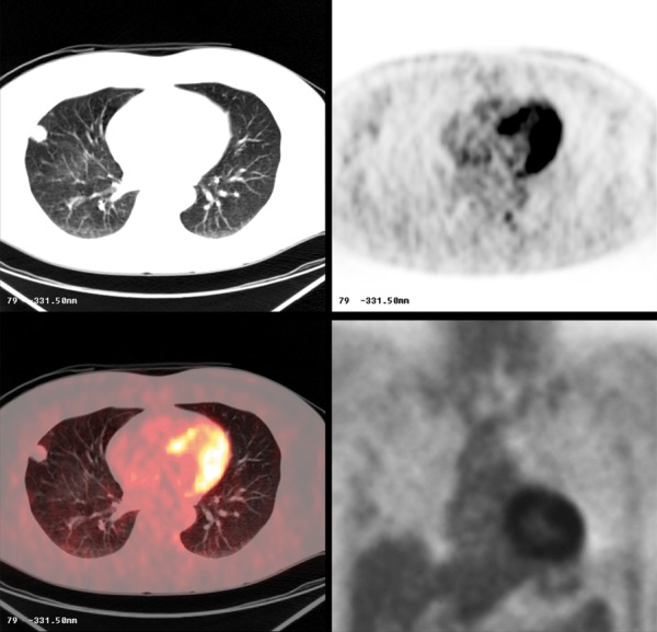
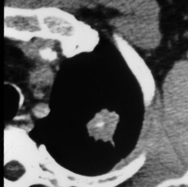
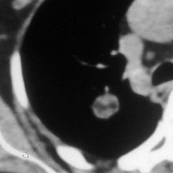
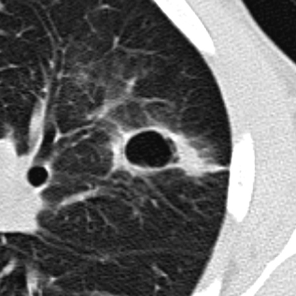
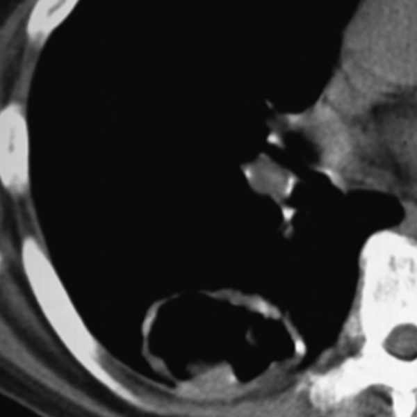
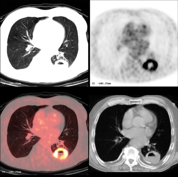
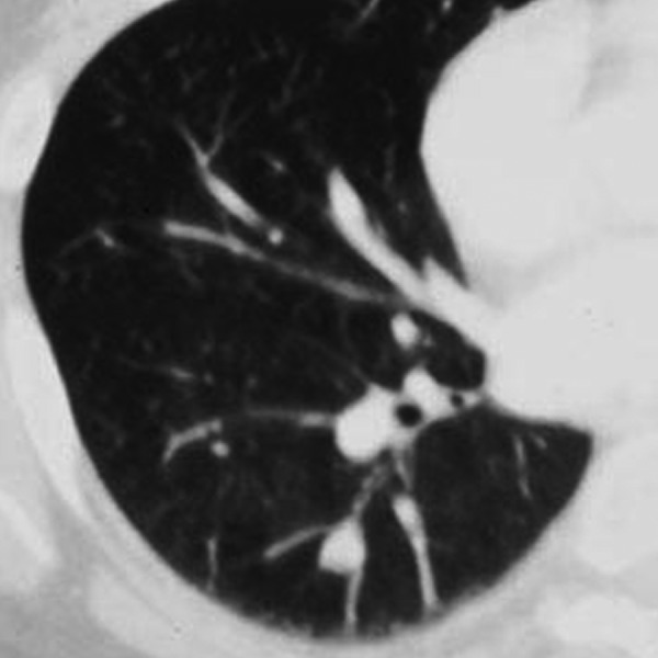
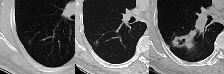
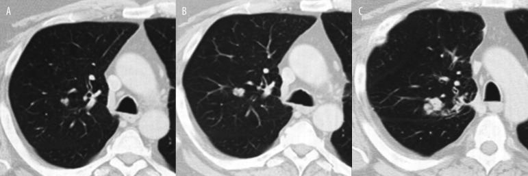
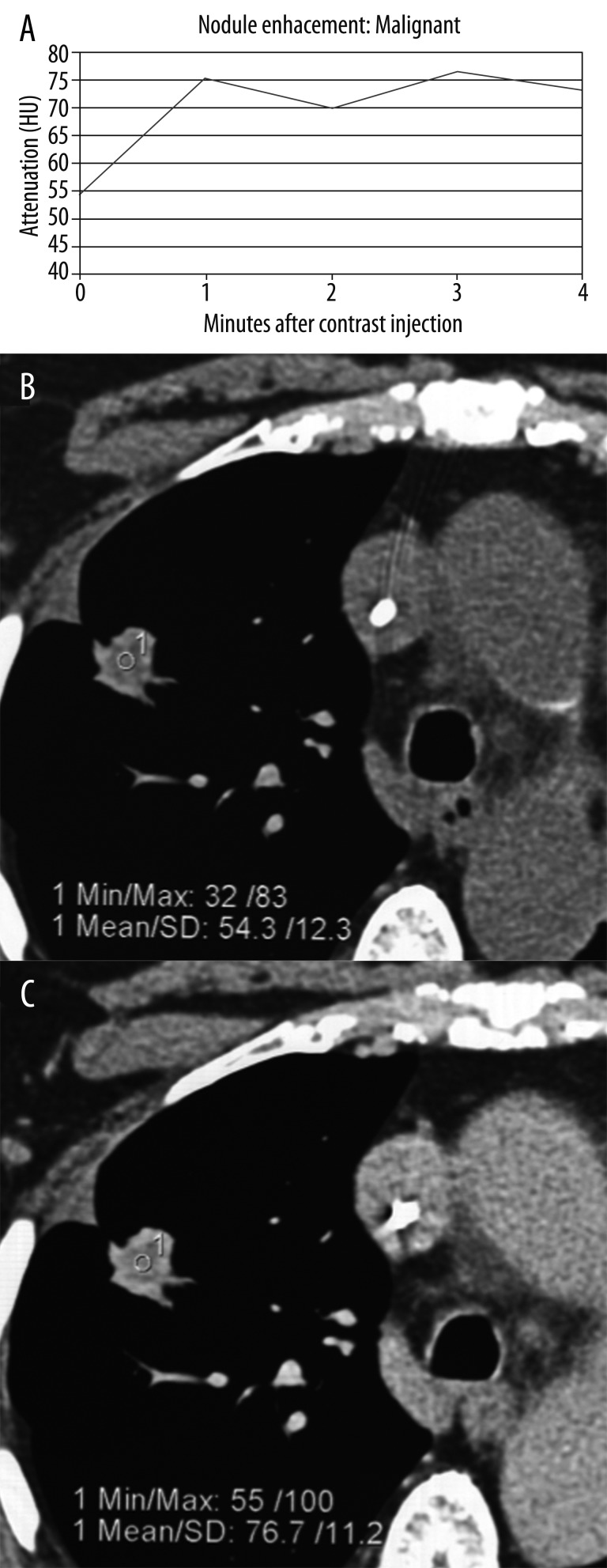
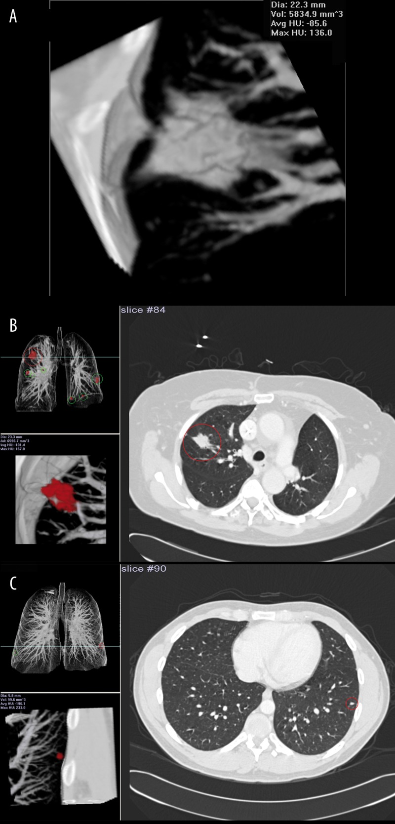
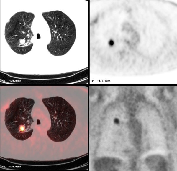
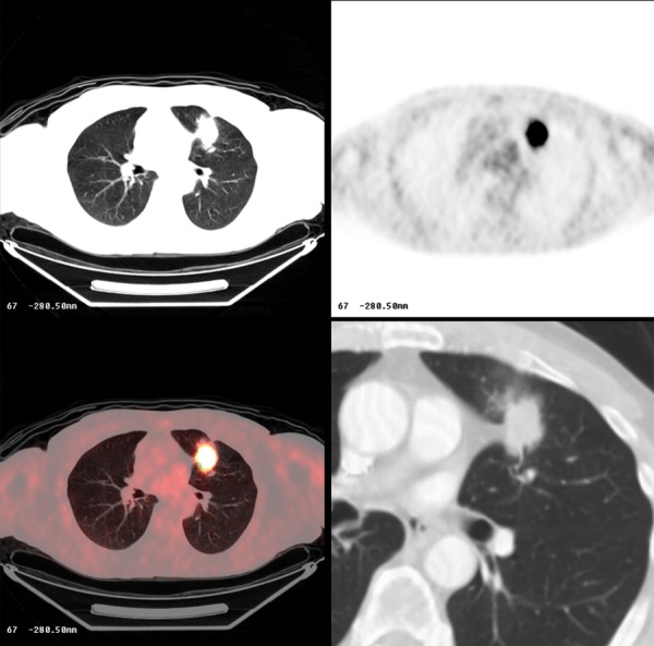
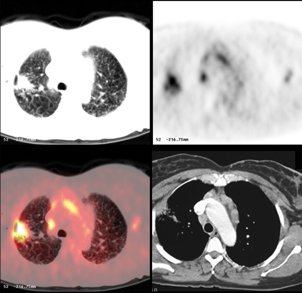
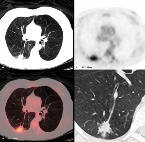
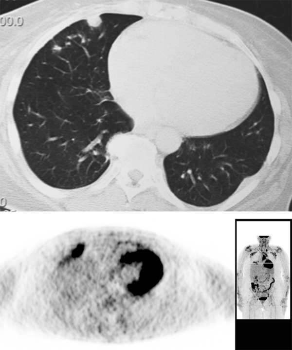
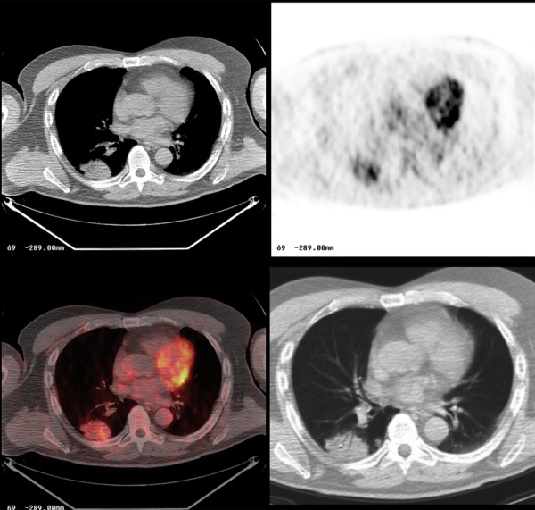
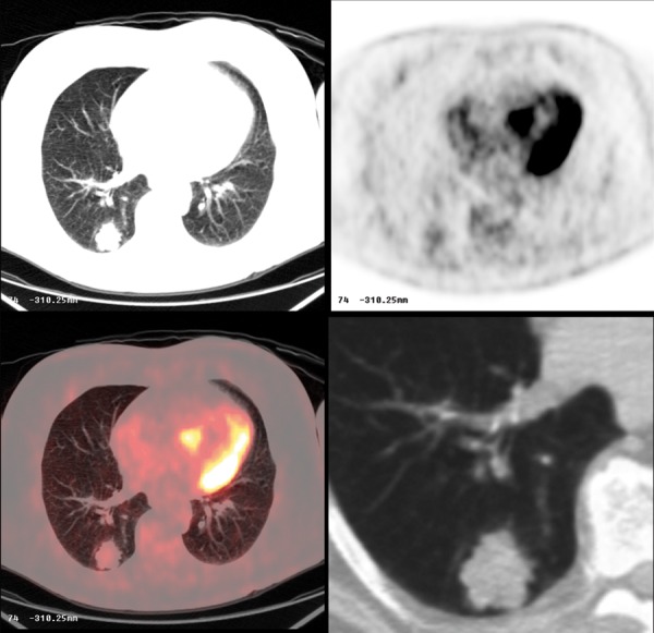
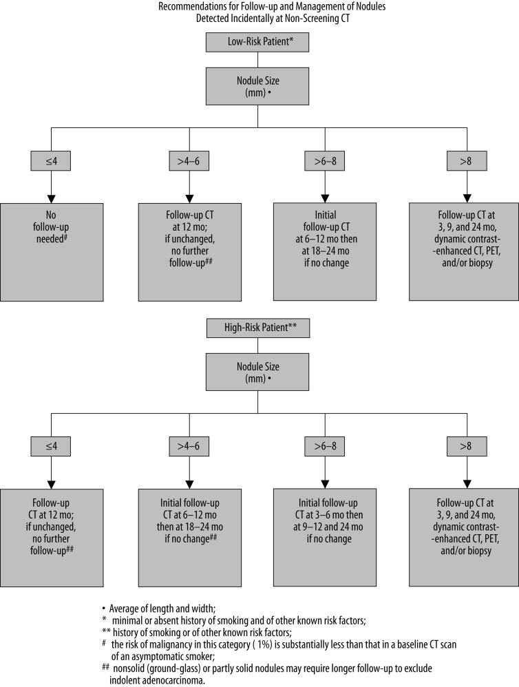
The solitary pulmonary nodule (SPN) has always been a diagnostic challenge for the radiologists. Currently, with increased utilization of computed tomography (CT) greater number of nodules is being discovered, with numerous indeterminate lesions, which frequently cannot be immediately classified into benign or malignant category.In this article we review the imaging features of benign and malignant round opacities; we demonstrate currently used standards and also more advanced techniques that are helpful in evaluating SPNs such as contrast-enhanced CT, PET/CT imaging and also pathologic sampling with biopsy or surgical resection.We also summarize the methods of evaluating and managing SPNs based on the latest guidelines from the Fleischner Society and American College of Chest Physicians.

Keywords: indeterminate nodule; multi-detector computed tomography (MDCT); solitary pulmonary nodule (SPN); transthoracic needle biopsy (TNB).


Figures

Figure 1
Figure 1
The differential diagnosis for a solitary pulmonary nodule [3,4].
Figure 2
Figure 2
Well-circumscribed pulmonary nodule with smooth margins.
Figure 3
Figure 3
Pulmonary nodule with irregular margins.
Figure 4
Figure 4
Pulmonary nodule with lobulated margins.
Figure 5
Figure 5
Pulmonary nodule with spiculated margins and „halo sign”.
Figure 6
Figure 6
Benign granuloma with characteristic laminated pattern of calcification.
Figure 7
Figure 7
Benign pulmonary nodule with central calcification due to prior histoplasma infection.
Figure 8
Figure 8
Histoplasmosis. Pulmonary nodule in CT and PET-CT. Note that the nodule is non FDG avid.
Figure 9
Figure 9
Pulmonary nodule with malignant pattern of calcification.
Figure 10.
Figure 10.
Benign hamartoma.
Figure 11
Figure 11
Benign cavity with relatively smooth, thin walls. This infectious lesion nearly resolved on follow-up imaging.
Figure 12
Figure 12
Metastatic cavitary nodule due to squamous cell carcinoma.
Figure 13
Figure 13
Cavitary squamous cell carcinoma. Note ring-like FDG avid lesion on PET-CT.
Figure 14
Figure 14
Carcinoid.
Figure 15
Figure 15
Slowly growing malignancy from a ground glass nodule to a cavitary mass over 7 years: (A). 5 mm at baseline, (B) 10 mm 3 years later, (C) cavitary mass at diagnosis 4 years later – poorly differentiated sarcomatoid carcinoma.
Figure 16
Figure 16
Moderately differentiated lung carcinoma: (A) baseline, (B) 10 months later, (C) 6 months later.
Figure 17
Figure 17
Pattern of malignant enhancement: (A) plot of nodule enhancement over time, (B) attenuation of the nodule before contrast administration, and (C) post-contrast enhancement.
Figure 18
Figure 18
Computer-aided detection/diagnosis (CAD) system R2: (A) volume calculation of the nodule, (B) system detected spiculated mass in the right lung, (C) tiny nodule detected in the left lung.
Figure 19
Figure 19
Moderately-differentiated lung carcinoma with FDG-avid lesion on PET-CT.
Figure 20
Figure 20
Poorly-differentiated lung carcinoma with FDG-avid lesion on PET-CT.
Figure 21
Figure 21
FDG avid inflammatory lesion in the right lung and mediastinal and hilar adenopathy in pulmonary sarcoidosis.
Figure 22
Figure 22
Aspergilloma in the right lung with FDG avid lesion on PET-CT.
Figure 23
Figure 23
Peripheral pulmonary nodule due to rheumatoid arthritis. Note FDG avid inflammatory nodule on PET.
Figure 24
Figure 24
Bronchioalveolar carcinoma in the right lung. Note mild, heterogeneous FDG avidity on PET-CT.
Figure 25
Figure 25
Metastatic pulmonary nodule from a colorectal mucinous adenocarcinoma. Note minimal FDG avidity on PET-CT.
Figure 26
Figure 26
The Fleischner Society recommendations for a follow-up and management of a solitary pulmonary nodule detected incidentally during a non-screening CT [55].

Similar articles

Cited by

References

    1. Austin JH, Muller NL, Friedman PJ, et al. Glossary of terms for CT of the lungs: Recommendations of the nomenclature committee of the Fleischner Society. Radiology. 1996;200:327–31. - PubMed
    1. Gould MK, Fletcher J, Iannettoni MD, et al. American College of Chest Physician. Evaluation of patients with pulmonary nodules: when is it lung cancer?: ACCP evidence-based clinical practice guidelines (2nd edition) Chest. 2007;132(Suppl.3):108S–30S. - PubMed
    1. Erasmus JJ, Connolly JE, McAdams HP, et al. Solitary pulmonary nodules: Part I. Morphologic evaluation for differentiation of benign and malignant lesions. Radiographics. 2000;20:43–58. - PubMed
    1. Brandman S, Ko JP. Pulmonary nodule detection, characterization, and management with multidetector computed tomography. J Thorac Imaging. 2011;26(2):90–105. - PubMed
    1. Nicholas E, Braff S, Klein JS. Evaluation of the solitary pulmonary nodule: A practical approach. Applied Radiology. http://www.appliedradiology.com/Issues/2011/12/Articles/AR_12-11_Klein-N... (December 2011; vol.40, number 12) (accessed 29.04.2012)

LinkOut - more resources