

Pneumatosis cystoides interstitialis: A complication of graft-versus-host disease. A report of two cases
- PMID: 22844311
- PMCID: PMC3403803
- DOI: 10.12659/pjr.882972
Pneumatosis cystoides interstitialis: A complication of graft-versus-host disease. A report of two cases
Abstract
Background: Pneumatosis cystoides intestinalis (PCI) is a rare disorder characterized by the presence of multiple gas collections in the subserosal or submucosal intestinal wall of the large or small intestine. We report two cases of PCI in the course of chronic graft-versus-host disease.
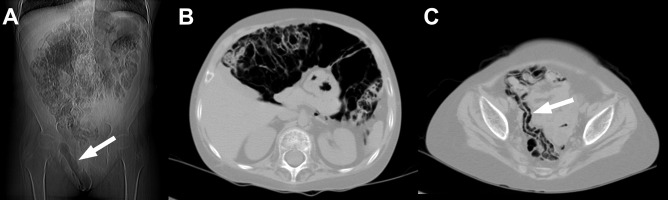
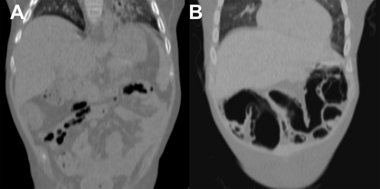
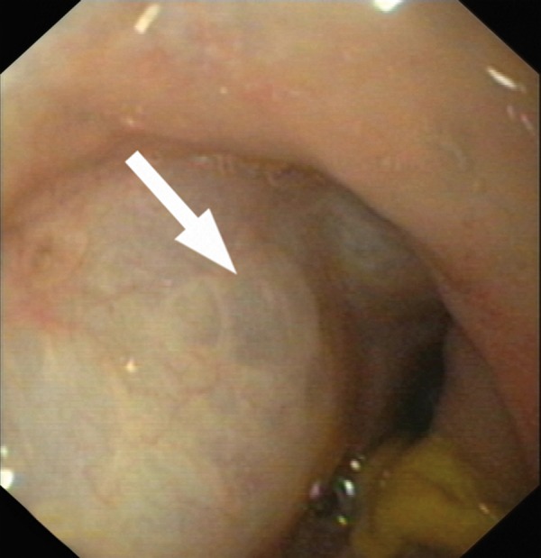
Case report: A 5-year-old girl was treated for acute lymphoblastic leukemia. Twenty-four months after the hematopoietic stem cell transplantation, in the course of graft-versus-host disease, she developed subcutaneous emphysema of the right inguinal and pudendal region. PCI was diagnosed based on a CT examination. A 3-year-old boy was treated for juvenile myelomonocytic leukemia. Fourteen months after the hematopoietic stem cell transplantation he presented with an increased severity of intestinal symptoms, including intermittent bleeding from large intestine. PCI was diagnosed based on a CT exam and was confirmed by a colonoscopy.
Conclusions: Pneumatosis cystoides interstitialis in the course of chronic graft-versus-host disease has a heterogeneous clinical presentation that does not correlate with results of imaging. CT is a method of choice to diagnose PCI. In patients with PCI, the presence of free air in the peritoneal cavity does not confirm an intestinal perforation.
Keywords: computed tomography; graft-versus-host disease; pneumatosis cystoides intestinalis.
Figures
Similar articles
-
Pneumatosis cystoides intestinalis: A rare complication after hematopoietic stem cell transplantation.Pediatr Transplant. 2021 Nov;25(7):e14136. doi: 10.1111/petr.14136. Epub 2021 Sep 10. Pediatr Transplant. 2021. PMID: 34505744
-
Acute lymphoblastic leukemia with pneumatosis cystoides intestinalis after hematopoietic stem cell transplantation: a case report.J Int Med Res. 2024 Sep;52(9):3000605241274581. doi: 10.1177/03000605241274581. J Int Med Res. 2024. PMID: 39246070 Free PMC article.
-
[Pneumatosis cystoides intestinalis developing subsequent to allogeneic hematopoietic stem cell transplantation for mixed phenotype acute leukemia].Rinsho Ketsueki. 2020;61(4):312-317. doi: 10.11406/rinketsu.61.312. Rinsho Ketsueki. 2020. PMID: 32378572 Japanese.
-
Pneumatosis cystoides intestinalis in scleroderma-related conditions.Intern Med J. 2012 Mar;42(3):323-9. doi: 10.1111/j.1445-5994.2011.02557.x. Intern Med J. 2012. PMID: 22432985 Review.
-
Pneumatosis cystoides intestinalis: case report and review of literature.Clin J Gastroenterol. 2020 Feb;13(1):31-36. doi: 10.1007/s12328-019-00999-3. Epub 2019 Jun 3. Clin J Gastroenterol. 2020. PMID: 31161540 Review.
Cited by
-
[Pneumatosis intestinalis after allogeneic hematopoietic stem cell transplantation: report of three cases and literature review].Zhonghua Xue Ye Xue Za Zhi. 2019 Mar 14;40(3):232-234. doi: 10.3760/cma.j.issn.0253-2727.2019.03.013. Zhonghua Xue Ye Xue Za Zhi. 2019. PMID: 30929392 Free PMC article. Chinese. No abstract available.
-
Spontaneous free perforation of the small intestine in adults.World J Gastroenterol. 2014 Aug 7;20(29):9990-7. doi: 10.3748/wjg.v20.i29.9990. World J Gastroenterol. 2014. PMID: 25110427 Free PMC article. Review.
-
Pneumatosis Cystoides Intestinalis.J Clin Diagn Res. 2017 Jun;11(6):TJ01-TJ02. doi: 10.7860/JCDR/2017/26197.10087. Epub 2017 Jun 1. J Clin Diagn Res. 2017. PMID: 28764267 Free PMC article. No abstract available.
-
Case Report: Massive Intestinal Pneumatosis and Pneumoretroperitoneum Following Hematopoietic Stem Cell Transplantation in a 2-Year-Old Child.Front Pediatr. 2021 Dec 8;9:700736. doi: 10.3389/fped.2021.700736. eCollection 2021. Front Pediatr. 2021. PMID: 34956969 Free PMC article.
-
Pneumatosis cystoides intestinalis, a rare case in a pediatric patient following allogeneic hematopoietic stem cell transplantation: CT findings and literature review.Radiol Case Rep. 2021 Aug 16;16(10):3120-3124. doi: 10.1016/j.radcr.2021.07.053. eCollection 2021 Oct. Radiol Case Rep. 2021. PMID: 34457100 Free PMC article.
References
Publication types
LinkOut - more resources
Full Text Sources
Miscellaneous