. 2012;7(7):e41298.
doi: 10.1371/journal.pone.0041298. Epub 2012 Jul 23.

Clonal analysis in recurrent astrocytic, oligoastrocytic and oligodendroglial tumors implicates IDH1- mutation as common tumor initiating event

Affiliations

Clonal analysis in recurrent astrocytic, oligoastrocytic and oligodendroglial tumors implicates IDH1- mutation as common tumor initiating event

Ulrike Lass et al. PLoS One. 2012.

Abstract

Background: To investigate the dynamics of inter- and intratumoral molecular alterations during tumor progression in recurrent gliomas.

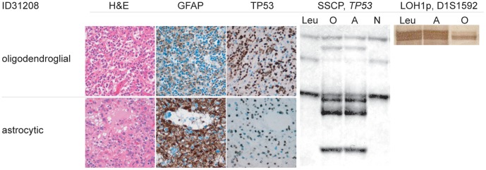
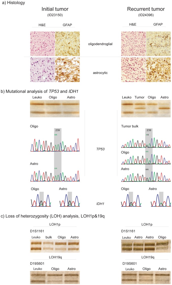
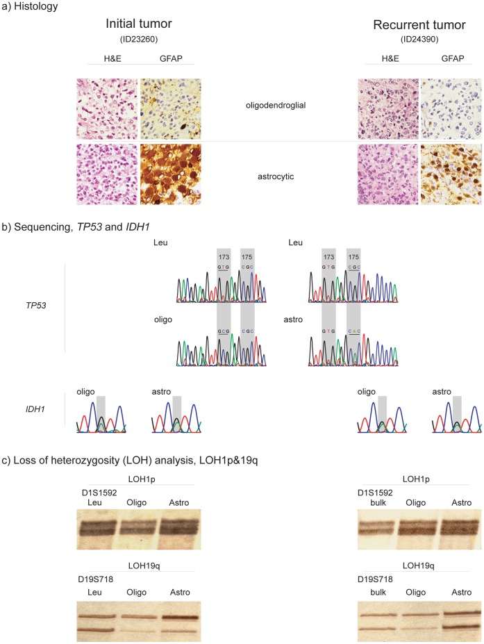
Methodology/principal findings: To address intertumoral heterogeneity we investigated non-microdissected tumor tissue of 106 gliomas representing 51 recurrent tumors. To address intratumoral heterogeneity a set of 16 gliomas representing 7 tumor pairs with at least one recurrence, and 4 single mixed gliomas were investigated by microdissection of distinct oligodendroglial and astrocytic tumor components. All tumors and tumor components were analyzed for allelic loss of 1p/19q (LOH 1p/19q), for TP53- mutations and for R132 mutations in the IDH1 gene. The investigation of non-microdissected tumor tissue revealed clonality in 75% (38/51). Aberrant molecular alterations upon recurrence were detected in 25% (13/51). 64% (9/14) of these were novel and associated with tumor progression. Loss of previously detected alterations was observed in 36% (5/14). One tumor pair (1/14; 7%) was significant for both. Intratumoral clonality was detected in 57% (4/7) of the microdissected tumor pairs and in 75% (3/4) of single microdissected tumors. 43% (3/7) of tumor pairs and one single tumor (25%) revealed intratumoral heterogeneity. While intratumoral heterogeneity affected both the TP53- mutational status and the LOH1p/19q status, all tumors with intratumoral heterogeneity shared the R132 IDH1- mutation as a common feature in both their microdissected components.

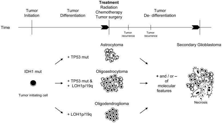
Conclusions/significance: The majority of recurrent gliomas are of monoclonal origin. However, the detection of divertive tumor cell clones in morphological distinct tumor components sharing IDH1- mutations as early event may provide insight into the tumorigenesis of true mixed gliomas.


Conflict of interest statement

Competing Interests: The authors have declared that no competing interests exist.

Figures

Figure 1
Figure 1. Histology, immunohistochemistry and molecular data of OIII, ID31208.
The tumor was significant for a small distinct tumor component of predominantly astrocytic appearance. At time of surgery this astrocytic component was considered too small to allow for the diagnosis of an OAIII. Molecular analysis detected an identical somatic TP-53- mutation in both the astrocytic and the oligodendroglial tumor component. However, LOH1p was detected in the oligodendroglial tumor component only. Both oligodendroglial and astrocytic tumor component shared the same IDH1- mutation (data not shown) suggesting a common tumor progenitor cell for both components. Upper panel (left three panels): Histology (H&E) and immunohistochemistry (GFAP, TP53) of the predominantly oligodendroglial tumor component. Lower panel (left three panels): Histology (H&E) and immunohistochemistry (GFAP, TP53) of the predominantly astrocytic tumor component. Of note, the oligodendroglial tumor component for the most part lacks GFAP, and nuclear accumulation of TP53- protein is seen in both tumor components. Middel panel: SSCP- mutational analysis of the predominantly oligodendroglial (O) and astrocytic (A) tumor component in comparison to patient’s leukocyte DNA (Leu) and a normal control (N). Identical aberrant bands were detected in both tumor components and later confirmed by direct sequencing. For reference see table 4. Right panel (LOH1p): Analysis of polymorphic micro satellite marker D1S1592 revealed retained alleles in the astrocytic tumor component (A). However, the oligodendroglial tumor component (O) was significant for the loss of one allele as compared to the patient’s leukocyte DNA (Leu).
Figure 2
Figure 2. Histology (H&E) and immunohistochemistry (GFAP) (a), SSCP and sequencing (b) and LOH1p/19q analyses (c) of OAII (ID23150, left panel, and its recurrence (ID24396), right panel.
(a) Histology Upper panel: H&E and GFAP of predominantly oligodendroglial tumor component in initial tumor, ID23150 (left) and its recurrence, ID24396 (right). Lower panel: H&E and GFAP of predominantly astrocytic tumor area in initial tumor, ID23150 (left) and its recurrence, ID24396 (right). The astrocytic tumor components in initial tumor and recurrence strongly stain positive for GFAP while the respective oligodendroglial tumor areas are almost devoid of GFAP staining. (b) Mutational analysis of TP53 and IDH1 Upper panel: SSCP- analysis of the micro-dissected oligodendroglial and predominantly astrocytic tumor component of the initial tumor, ID23159, (left) and non-microdissected and micro-dissected tumor areas in the recurrence, ID24396, (right). No aberrant band was detected in the initial tumor, as compared to the patient’s leukocyte DNA (Leuko). In the recurrent non-microdissected tumor tissue (tumor) and the micro-dissected astrocytic tumor component (Astro) an aberrantly shifted band was detected in exon 7 of the TP53- gene. Of note, the aberrant band was confined to the astrocytic tumor component. The oligodendroglial tumor part (Oligo) was significant for wild type TP53. Middle panel: Direct sequencing of the TP53-gene, exon 7 in micro-dissected oligodendroglial (Oligo) and astrocytic (Astro) tumor components in the initial tumor, ID23150 (left) and the non-microdissected tissue and the respective micro-dissected areas in the recurrent tumor, ID24396 (right). Sequencing confirms SSCP data for both the initial and the recurrent tumor. While the initial tumor is wild-type- TP53 in both oligodendroglial and astrocytic tumor component, the non-microdissected tissue of the recurrent tumor reveals heterozygosity for a somatic A->G point mutation in codon239 of the TP53- gene. Consequently amino acid aspartate (D) is substituted by asparagine (N). For reference see also table 4. The somatic mutation was confined to the astrocytic tumor component (Astro). The oligodendroglial tumor part remained wild-type- TP53 (Oligo), suggesting two distinct tumor cell clones. Lower panel: Direct sequencing of IDH1- gene. Identical G395A mutations in codon 132 (R132H) of IDH1 were detected in both oligodendroglial and astrocytic tumor components. (c) Loss of heterozygosity (LOH) analysis, LOH1p/19q Upper panel: LOH1p- analysis of polymorphic microsatellite marker (D1S1161) of the micro-dissected initial tumor, ID23150 (left) and its recurrence, ID24396 (right). The initial tumor and its recurrence are significant for LOH1p of the oligodendroglial tumor component only (Oligo). Both alleles were retained in the predominantly astrocytic tumor part (Astro) and the non-microdissected tissue for both the initial (left) and the recurrent tumor (right), again indicating two distinct tumor cell populations. Lower panel: LOH19q- analysis of polymorphic microsatellite marker (D19S601) of the micro-dissected initial tumor, ID23150 (left) and its recurrence, ID24396 (right). Both oligodendroglial (Oligo) and astrocytic (Astro) tumor component of initial and recurrent tumor revealed LOH19q. Patient’s leukocyte DNA (Leuko) was used for reference for both LOH1p- and LOH19q- analyses.
Figure 3
Figure 3. Histology (H&E) and immunohistochemistry (GFAP) (a), sequencing (b) and LOH1p/19q analyses (c) of OAIII (ID23260), left panel, and its recurrence (ID24390), right panel.
(a) Histology Upper panel: H&E and GFAP of predominantly oligodendroglial tumor component in initial tumor, ID23260 (left) and its recurrence, ID24390 (right). Lower panel: H&E and GFAP of predominantly astrocytic tumor area in initial tumor, ID23260 (left) and its recurrence, ID24390 (right). The astrocytic tumor components in initial tumor and recurrence strongly stain positive for GFAP while the respective oligodendroglial tumor areas are almost devoid of GFAP- staining. (b) Sequencing, TP53 and IDH1 Upper panel: Direct sequencing of TP53- gene, exon 5 of the patient’s leukocyte DNA (Leu) revealing wild- type sequence. Lower panel: Direct sequencing of TP53- gene, exon 5 of microdissected intial and recurrent tumor. Of note, the oligodendroglial tumor component harboured a point mutation in codon 173 of the TP53- gene and an amino acid substitution of valine -> alanine, while the astrocytic tumor component was significant for a point mutation in codon 175 of the same gene, resulting in an aminoacid substitution arginine -> histidine. Both initial and recurrent tumor harboured the same somatic TP53- mutations in the distinct tumor parts, confirming two distinct tumor cell populations in two independent analyses. For reference see also table 4. Lower panel: Direct sequencing of IDH1- gene. Identical G395A mutations in codon 132 (R132H) of IDH1 were detected in oligodendroglial and astrocytic tumor components of both the primary tumors and its related recurrence. (c) Upper panel: LOH1p- analysis of polymorphic microsatellite marker (D1S1592) of the micro-dissected initial tumor, ID22360 (left) and its recurrence, ID24390 (right). The initial tumor and its recurrence are significant for LOH1p of the oligodendroglial tumor component only (Oligo). Both alleles were retained in the predominantly astrocytic tumor part (Astro) in both the initial (left) and the recurrent tumor (right), indicating two distinct tumor cell populations. Lower panel: LOH19q- analysis of polymorphic microsatellite marker (D19S718) of the micro-dissected initial tumor, ID23260 (left) and its recurrence, ID24390 (right). Both oligodendroglial (Oligo) and astrocytic (Astro) tumor component of initial tumor revealed LOH19q. While LOH19q was still detectable in the oligodendroglial tumor component (Oligo) of the recurrent tumor, both alleles were retained in the recurrent astrocytic tumor part, supporting the existence of independent tumor subclones. Patient’s leukocyte DNA (Leuko) was used for reference for both LOH1p- and LOH19q- analyses.
Figure 4
Figure 4. Potential hierarchy of molecular events during tumor initiation, differentiation and de-differentiation upon tumor recurrence in pure and mixed gliomas.
Time line of tumor development from left to right. Tumor initiation occurs by the introduction of an IDH1- mutation in a common tumor progenitor cell. Tumor differentiation is significant for astrocytoma- typical molecular alterations in pure astrocytomas (i.e. TP53- mutations) and oligodendroglioma- typical alterations in pure oligodendrogliomas (i.e. LOH1p&19q). Of note, in pure oligoastrocytomas, TP53- mutations and LOH1p&19q may occur separately in the tumor parts with the respective morphology (oligodendroglial vs. astrocytic). Following tumor therapy with surgery, radio- and chemotherapy additional molecular events may occur at tumor recurrence. Also, previous molecular alterations might disappear due to overgrowth of therapy- resistant tumor clones or the disappearance of tumor- susceptible tumor cell clones. Tumor dedifferentiation finally leads to the morphological picture of a secondary glioblastoma with or without an oligodendroglial component as the common morphological endstage of malignant gliomas.

References

    1. Cairncross JG, Ueki K, Zlatescu MC, Lisle DK, Finkelstein DM, et al. Specific genetic predictors of chemotherapeutic response and survival in patients with anaplastic oligodendrogliomas. J Natl Cancer Inst. 1998;90:1473–1479. - PubMed
    1. Smith JS, Perry A, Borell TJ, Lee HK, O'Fallon J, et al. Alterations of chromosome arms 1p and 19q as predictors of survival in oligodendrogliomas, astrocytomas, and mixed oligoastrocytomas. J Clin Oncol. 2000;18:636–645. - PubMed
    1. Felsberg J, Erkwoh A, Sabel MC, Kirsch L, Fimmers R, et al. Oligodendroglial tumors: refinement of candidate regions on chromosome arm 1p and correlation of 1p/19q status with survival. Brain Pathol. 2004;14:121–130. - PMC - PubMed
    1. Giannini C, Burger PC, Berkey BA, Cairncross JG, Jenkins RB, et al. Anaplastic oligodendroglial tumors: refining the correlation among histopathology, 1p 19q deletion and clinical outcome in Intergroup Radiation Therapy Oncology Group Trial 9402. Brain Pathol. 2008;18:360–369. - PMC - PubMed
    1. Hegi ME, Diserens AC, Gorlia T, Hamou MF, de Tribolet N, et al. MGMT gene silencing and benefit from temozolomide in glioblastoma. N Engl J Med. 2005;352:997–1003. - PubMed

Publication types

MeSH terms