. 2012;7(7):e41299.
doi: 10.1371/journal.pone.0041299. Epub 2012 Jul 23.

Estrogens induce expression of membrane-associated estrogen receptor α isoforms in lactotropes

Affiliations

Estrogens induce expression of membrane-associated estrogen receptor α isoforms in lactotropes

Sandra Zárate et al. PLoS One. 2012.

Abstract

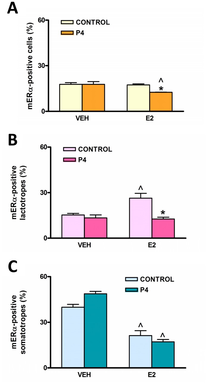
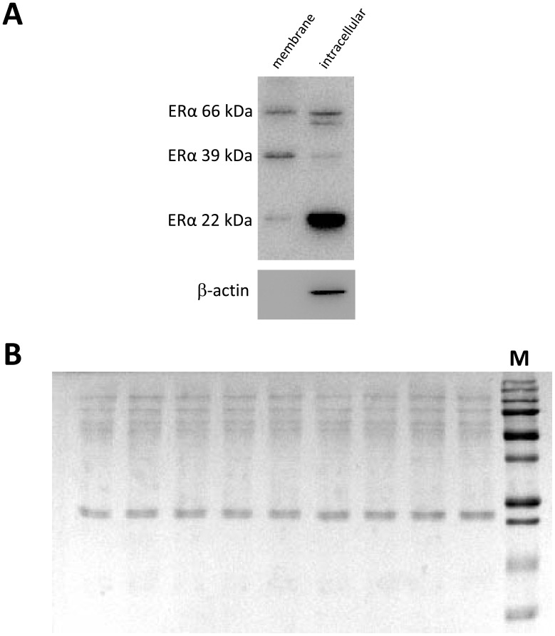
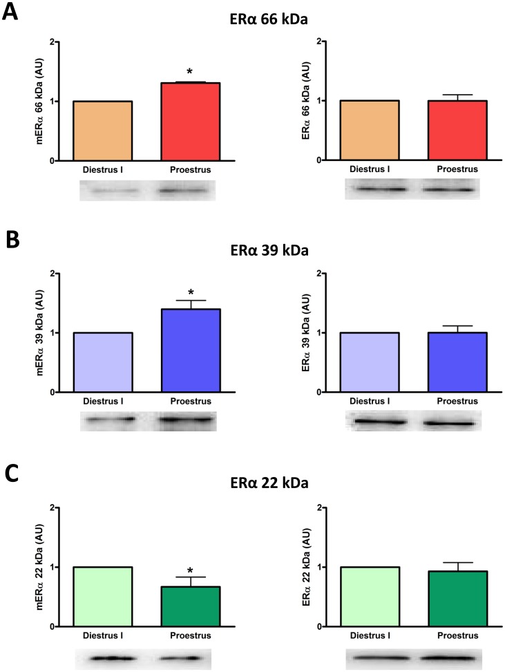
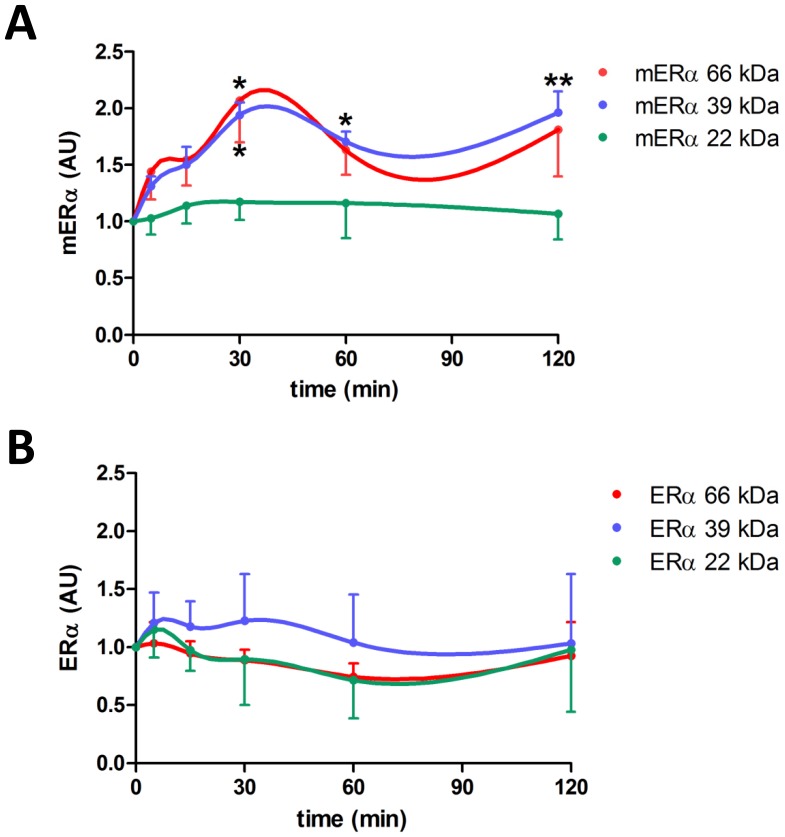
Estrogens are key to anterior pituitary function, stimulating hormone release and controlling cell fate to achieve pituitary dynamic adaptation to changing physiological conditions. In addition to their classical mechanism of action through intracellular estrogen receptors (ERs), estrogens exert rapid actions via cell membrane-localized ERs (mERs). We previously showed that E2 exerts a rapid pro-apoptotic action in anterior pituitary cells, especially in lactotropes and somatotropes, through activation of mERs. In the present study, we examined the involvement of mERα in the rapid pro-apoptotic action of estradiol by TUNEL in primary cultures of anterior pituitary cells from ovariectomized rats using a cell-impermeable E2 conjugate (E2-BSA) and an ERα selective antagonist (MPP dihydrochloride). We studied mERα expression during the estrous cycle and its regulation by gonadal steroids in vivo by flow cytometry. We identified ERα variants in the plasma membrane of anterior pituitary cells during the estrous cycle and studied E2 regulation of these mERα variants in vitro by surface biotinylation and Western Blot. E2-BSA-induced apoptosis was abrogated by MPP in total anterior pituitary cells and lactotropes. In cycling rats, we detected a higher number of lactotropes and a lower number of somatotropes expressing mERα at proestrus than at diestrus. Acute E2 treatment increased the percentage of mERα-expressing lactotropes whereas it decreased the percentage of mERα-expressing somatotropes. We detected three mERα isoforms of 66, 39 and 22 kDa. Expression of mERα66 and mERα39 was higher at proestrus than at diestrus, and short-term E2 incubation increased expression of these two mERα variants. Our results indicate that the rapid apoptotic action exerted by E2 in lactotropes depends on mERα, probably full-length ERα and/or a 39 kDa ERα variant. Expression and activation of mERα variants in lactotropes could be one of the mechanisms through which E2 participates in anterior pituitary cell renewal during the estrous cycle.


Conflict of interest statement

Competing Interests: The authors have declared that no competing interests exist.

Figures

Figure 1
Figure 1. An ERα-antagonist (MPP) blocks E2-BSA-induced apoptosis of lactotropes.
Anterior pituitary cells in culture were pre-incubated with MPP (100 nM) for 30 min prior to the addition of E2-BSA (1 nM) or VEH for 120 min. Apoptosis was analyzed by TUNEL and lactotropes and somatotropes were identified by immunocytochemistry. Each column represents the percentage ± CI (95%) of TUNEL-positive cells (n≥2600) (A), lactotropes (n≥1500 cells/group) (B) and somatotropes (n≥1100 cells/group) (C). *p<0.05 vs respective control without MPP; ∧p<0.05, formula image
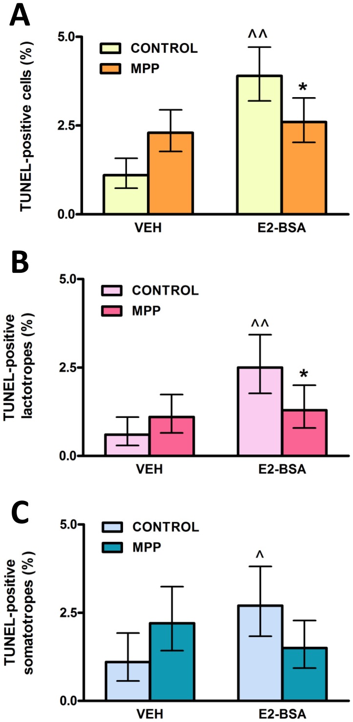
<0.01 vs respective control without E2-BSA, χ2 test.
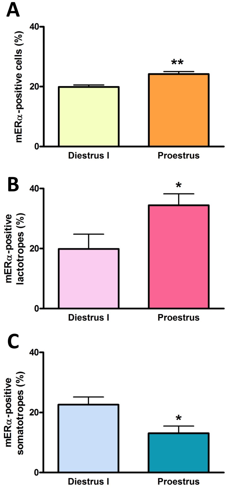
Figure 2
Figure 2. The percentage of lactotropes and somatotropes expressing mERα varies during the estrous cycle.
Dispersed anterior pituitary cells from rats euthanized at diestrus I or proestrus were immunostained for both mERα and PRL or GH and analyzed by flow cytometry. Each column represents the mean ± SE of anterior pituitary cells (A), lactotropes (B) or somatotropes (C) expressing mERα (n = 3–4 animals per group). *p<0.05, **p<0.01 vs diestrus I, Student’s t test.
Figure 3
Figure 3. Gonadal steroids differentially regulate the expression of mERα in lactotropes and somatotropes in vivo.
OVX female rats were injected for two consecutive days with vehicle (VEH), 17β-estradiol (E2), progesterone (P4) or E2+P4 and euthanized on the third day. Dispersed anterior pituitary cells were immunostained for both mERα and PRL or GH and analyzed by flow cytometry. Each column represents the mean ± SE of anterior pituitary cells (A), lactotropes (B) or somatotropes (C) expressing mERα (n = 3−4 animals per group). *p<0.05 vs respective control without P4; ∧p<0.05 vs respective control without E2, two-way ANOVA followed by Tukey’s test.
Figure 4
Figure 4. Cell surface biotinylation of anterior pituitary cells.
Cultured anterior pituitary cells from cycling rats were processed for cell surface biotinylation as described in Material and Methods. Western blots from biotinylated (membrane) and non-biotinylated (intracellular) protein fractions were probed with anti-rat ERα and β-actin antibodies (A). PVDF membranes from gels loaded with membrane protein fractions were stained with reversible Ponceau S to ensure equal loading of proteins (B). M: MW marker.
Figure 5
Figure 5. The expression of mERα isoforms varies during the estrous cycle.
Anterior pituitary cells from cycling female rats killed at diestrus I or proestrus were cultured for 24 h to allow attachment to the culture plate and then processed for cell surface biotinylation. Expression of full-length ERα (A) and its 39 kDa (B) and 22 kDa (C) isoforms was evaluated by Western blot in membrane (left panels) and intracellular (right panels) protein fractions. Densitometric data from 3–6 animals per group were normalized by the corresponding Ponceau staining (left panels) or β-actin value (right panels) and analyzed by paired Student’s t test, *p<0.05 vs diestrus I. Each column represents the mean ± SE of the relative increment of proestrus versus corresponding diestrus I.
Figure 6
Figure 6. 17β-estradiol increases expression of 66 kDa (full-length) and 39 kDa mERα isoforms in a time-dependent manner.
Anterior pituitary cells from OVX rats in culture were incubated with E2 (1 nM) for 0–120 min and then processed for cell surface biotinylation. Expression of full-length ERα and its isoforms was evaluated by Western Blot in membrane (A) and intracellular (B) protein fractions. Densitometric data from 3–5 experiments were normalized by the corresponding Ponceau staining (A) or β-actin value (B) and analyzed by repeated measures one-way ANOVA followed by Dunnett’s multiple comparisons test, *p<0.05, **p<0.01 vs 0 min. Each point represents the mean ± SE of the relative increment of each time compared to corresponding time 0 min for each ERα isoform.

References

    1. Shupnik MA. Oestrogen receptors, receptor variants and oestrogen actions in the hypothalamic-pituitary axis. J Neuroendocrinol. 2002;14:85–94. - PubMed
    1. Lewis-Wambi JS, Jordan VC. Estrogen regulation of apoptosis: how can one hormone stimulate and inhibit? Breast Cancer Res. 2009;11:206. - PMC - PubMed
    1. Jensen EV, Jacobson HI. Basic guides to the mechanism of estrogen action. Rec Prog Horm Res. 1962;18:387–414.
    1. McDevitt MA, Glidewell-Kenney C, Jimenez MA, Ahearn PC, Weiss J, et al. New insights into the classical and non-classical actions of estrogen: evidence from estrogen receptor knock-out and knock-in mice. Mol Cell Endocrinol. 2008;290:24–30. - PMC - PubMed
    1. Madak-Erdogan Z, Kieser KJ, Kim SH, Komm B, Katzenellenbogen JA, et al. Nuclear and extranuclear pathway inputs in the regulation of global gene expression by estrogen receptors. Mol Endocrinol. 2008;22:2116–27. - PMC - PubMed

Publication types