. 2012;7(7):e41399.
doi: 10.1371/journal.pone.0041399. Epub 2012 Jul 23.

β-Glucan derived from Aureobasidium pullulans is effective for the prevention of influenza in mice

Affiliations

β-Glucan derived from Aureobasidium pullulans is effective for the prevention of influenza in mice

Daisuke Muramatsu et al. PLoS One. 2012.

Abstract

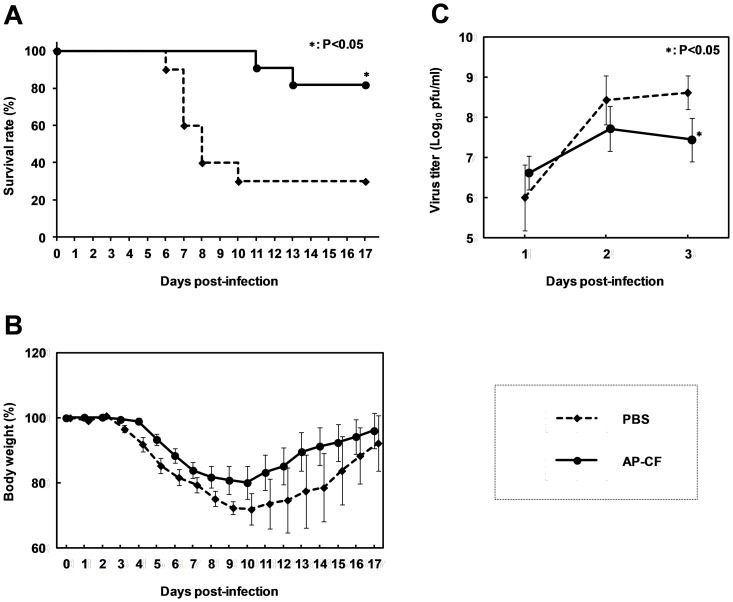
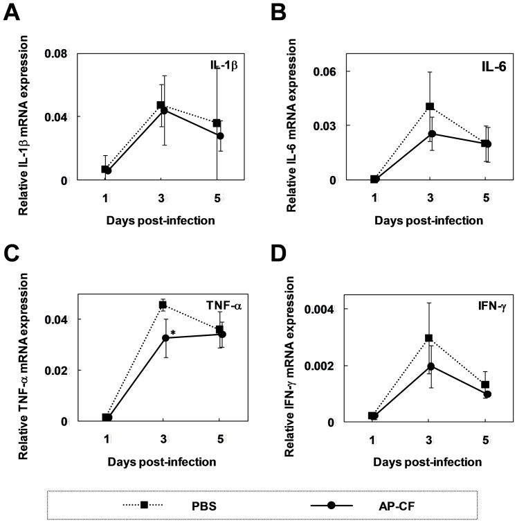
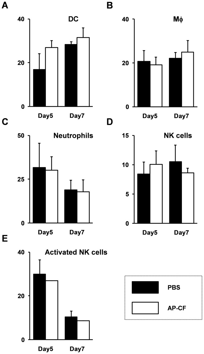
β-(1→3)-D-glucans with β-(1→6)-glycosidic linked branches produced by mushrooms, yeast and fungi are known to be an immune activation agent, and are used in anti-cancer drugs or health-promoting foods. In this report, we demonstrate that oral administration of Aureobasidium pullulans-cultured fluid (AP-CF) enriched with the β-(1→3),(1→6)-D-glucan exhibits efficacy to protect mice infected with a lethal titer of the A/Puerto Rico/8/34 (PR8; H1N1) strain of influenza virus. The survival rate of the mice significantly increased by AP-CF administration after sublethal infection of PR8 virus. The virus titer in the mouse lung homogenates was significantly decreased by AP-CF administration. No significant difference in the mRNA expression of inflammatory cytokines, and in the population of lymphocytes was observed in the lungs of mice administered with AP-CF. Interestingly, expression level for the mRNA of virus sensors, RIG-I (retinoic acid-inducible gene-I) and MDA5 (melanoma differentiation-associated protein 5) strongly increased at 5 hours after the stimulation of A. pullulans-produced purified β-(1→3),(1→6)-D-glucan (AP-BG) in murine macrophage-derived RAW264.7 cells. Furthermore, the replication of PR8 virus was significantly repressed by pre-treatment of AP-BG. These findings suggest the increased expression of virus sensors is effective for the prevention of influenza by the inhibition of viral replication with the administration of AP-CF.


Conflict of interest statement

Competing Interests: Mr. Yasuhiro Nikawa and Dr. Mitsuyasu Okabe are employees of Aureo Co., Ltd., and Mr. Daisuke Muramatsu, Dr. Atsushi Iwai, Miss Shiho Aoki, Mr. Hirohumi Uchiyama, Dr. Koji Kawata, and Mr. Yasuhiro Nikawa are employees of Aureo-Science Co., Ltd. The results presented in this manuscript are patent pending in Japan (application number: 2009-234023). There are no other patents, products in development or marketed products to declare. This does not alter the authors' adherence to all the PLoS ONE policies on sharing data and materials.

Figures

Figure 1
Figure 1. The oral administration of Aureobasidium pullulans-cultured fluid (AP-CF) protects mice from lethal PR8 virus infections.
(A, B) AP-CF (40 µg/mouse, n = 11), or PBS (n = 10) was orally administered to the C57BL/6N mice throughout the experiment once a day, from 7 days before the virus infection. After the 7 day pre-treatment, the mice were intranasally infected with the A/PuertoRico/8/34 (PR8; H1N1) strain of influenza A virus at a titer of 2,000 pfu, and then the survival rate (A) and the body weight loss (B) of the mice were recorded. Error bars indicate the standard error of the mean. (C) The mice (n = 5 for each group) were infected with influenza A virus at the same condition as in panel A and B, and then the lungs of the mice were collected at the day indicated in the figure. Virus titers in the lung homogenates were measured by plaque assay. The error bars indicate standard deviations which were calculated by five independent experiments. An asterisk (*) indicates P<0.05 compared with the mice treated with PBS.
Figure 2
Figure 2. The oral administration of AP-CF inhibits influenza A virus replication in the lung.
Total RNAs were isolated from the lung tissue of mice infected with influenza A virus at the day indicated in each panel. Subsequently, the RNAs were subjected to real-time RT-PCR analysis using a specific primer set for interleukin-1β (IL-1β; A), IL-6 (B), tumor necrosis factor-α (TNF-α; C), or interferon-γ (IFN-γ; D). The data represent relative mRNA expressions which were normalized with the expression level of glyceraldehyde-3-phosphate dehydrogenase (G3PDH) mRNA. Error bars indicate standard deviations which were calculated with three independent experiments. An asterisk (*) indicates P<0.05 compared with the control mice.
Figure 3
Figure 3. The population of lung immune cells except DC cells is not significantly affected by the oral administration of AP-CF after an influenza A virus infection.
The C57BL/6N mice were infected with the influenza A virus at the same condition as in Figure 1, and then the whole lung tissues were extracted from the mice. The cells were dissociated by the treatment with collagenase and DNaseI, then stained with fluorescent labeled specific antibodies and analyzed by the FACS (FACS Canto; BD bioscience). The data represent the percentage of the cells from total alive cells (7-AAD negative cells). Error bars indicate standard deviations which were calculated with three independent experiments. (A) dendritic cell (DC); CD11c+, F4/80; (B) macrophage (Mφ): F4/80+; (C) neutrophills: Gr-1+; (D) natural killer (NK) cell: CD49b+, CD3; (E) activated NK cell: CD49b+, CD3, CD69+.
Figure 4
Figure 4. Stimulation with purified Aureobasidium pullans-produced β-(1→3),(1→6)-D-glucan (AP-BG) induces the production of growth factors and inflammatory cytokines in RAW264.7 cells.
(A–E) RAW264.7 cells were stimulated with AP-BG in a concentration of 100 µg/ml, and then the cells were harvested at some time points of post-stimulation indicated in the figure. Total RNAs isolated from the cells were then analyzed by real-time RT-PCR using specific primer sets for interleukin-1β (IL-1ß; A), IL-6 (B), tumor necrosis factor-α (TNF-α; C), granulocyte-macrophage colony-stimulating factor (GM-CSF; D), and granulocyte colony-stimulating factor (G-CSF; E). The expression of genes was normalized with the expression of G3PDH mRNA, and the data represent a relative expression amount compared with the initial time point. Error bars indicate standard deviations which were calculated with three independent experiments. Single asterisk (*) and double asterisk (**) indicate significant differences between AP-BG-treated cells and control cells with P<0.05 and P<0.01, respectively.
Figure 5
Figure 5. Expression of RIG-I and MDA5 is upregulated in the RAW264.7 cells, after AP-BG stimulation.
(A–C) RAW264.7 cells were stimulated with AP-BG at the concentration of 100 µg/ml. At the time point indicated in the figure, the cells were harvested, and the total RNAs isolated from the cells were subjected to real-time RT-PCR analysis using specific primer sets for retinoic acid-inducible gene-I (RIG-I; A), melanoma differentiation-associated protein 5 (MDA5; B), and interferon-β promoter stimulator 1 (IPS-1; C). The data represent relative mRNA expression compared with the expression level of the initial time point, and the calculated values for each time point were normalized with the expression level of G3PDH mRNA. Error bars indicate standard deviations which were calculated by three independent experiments. (D, E) THP-1 cells which were differentiated into macrophages using 100 nM of phorbol 12-myristate 13-acetate (PMA), were stimulated with 100 µg/ml of AP-BG. After 6 hrs, the cells were harvested, and the total RNAs isolated from the cells were subjected to the real-time RT-PCR analysis using specific primer sets for RIG-I (D) and MDA5 (E). (F) RAW264.7 cells were stimulated with AP-BG (100 µg/ml) or PBS for 6 hrs, and then the PR8 strain of influenza A virus was infected to the cells (MOI = 10). After the incubation for the periods indicated in the figure, the virus titers in the cultured medium were measured by plaque assay. Single asterisk (*) and double asterisk (**) indicate significant differences between AP-BG-treated cells and control cells with P<0.05 and P<0.01, respectively.

References

    1. Akramiene D, Kondrotas A, Didziapetriene J, Kevelaitis E. Effects of beta-glucans on the immune system. Medicina (Kaunas) 2007;43:597–606. - PubMed
    1. Chan GC, Chan WK, Sze DM. The effects of beta-glucan on human immune and cancer cells. J Hematol Oncol. 2009;2:25. - PMC - PubMed
    1. Novak M, Vetvicka V. Glucans as biological response modifiers. Endocr Metab Immune Disord Drug Targets. 2009;9:67–75. - PubMed
    1. Chaung HC, Huang TC, Yu JH, Wu ML, Chung WB. Immunomodulatory effects of beta-glucans on porcine alveolar macrophages and bone marrow haematopoietic cell-derived dendritic cells. Vet Immunol Immunopathol. 2009;131:147–157. - PubMed
    1. Liu J, Gunn L, Hansen R, Yan J. Yeast-derived beta-glucan in combination with anti-tumor monoclonal antibody therapy in cancer. Recent Pat Anticancer Drug Discov. 2009;4:101–109. - PubMed

Publication types

MeSH terms