. 2012;7(7):e41445.
doi: 10.1371/journal.pone.0041445. Epub 2012 Jul 23.

Thyroid hormone enhances nitric oxide-mediated bacterial clearance and promotes survival after meningococcal infection

Affiliations

Thyroid hormone enhances nitric oxide-mediated bacterial clearance and promotes survival after meningococcal infection

Yao Chen et al. PLoS One. 2012.

Abstract

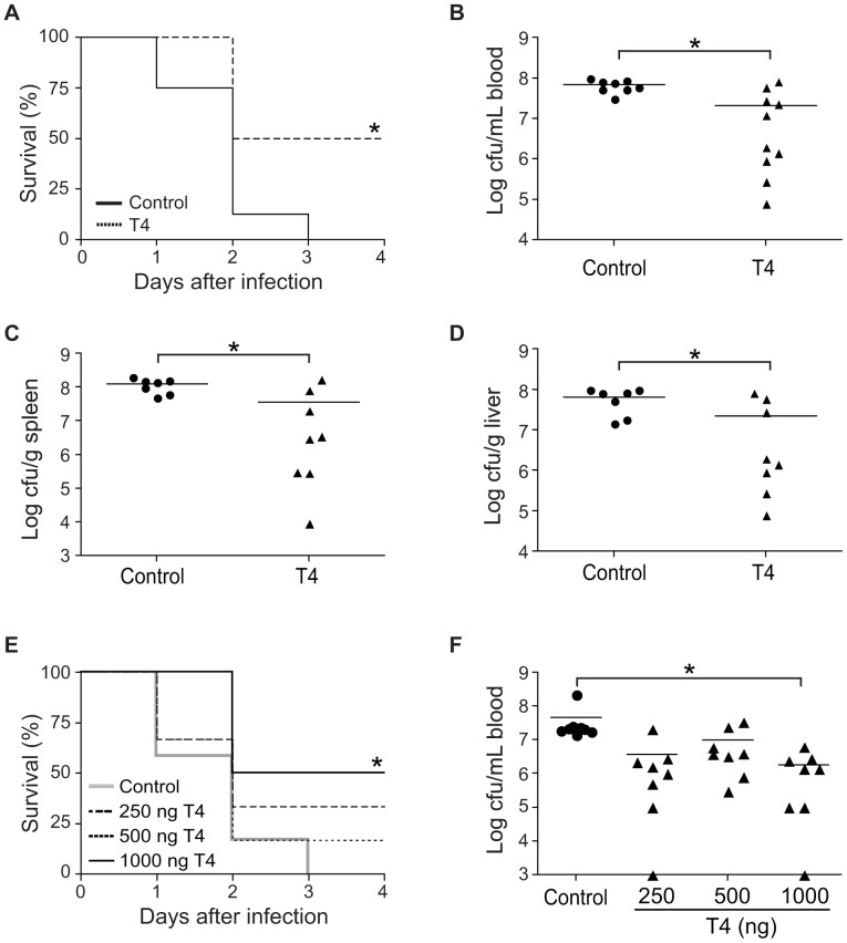
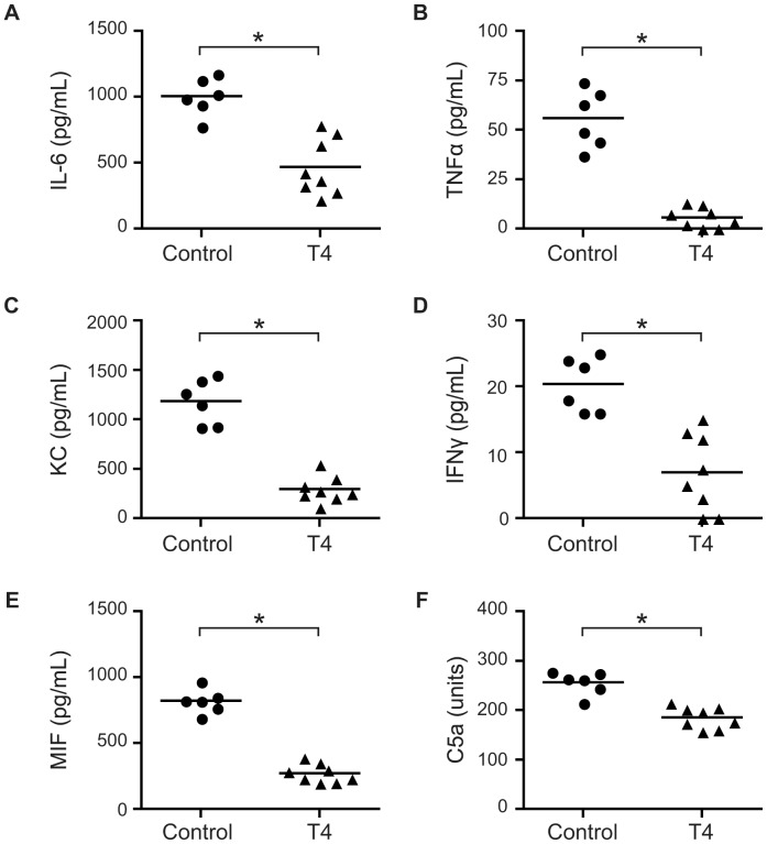
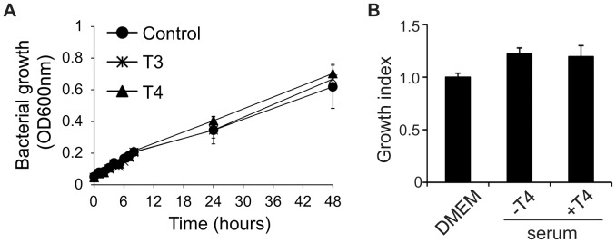
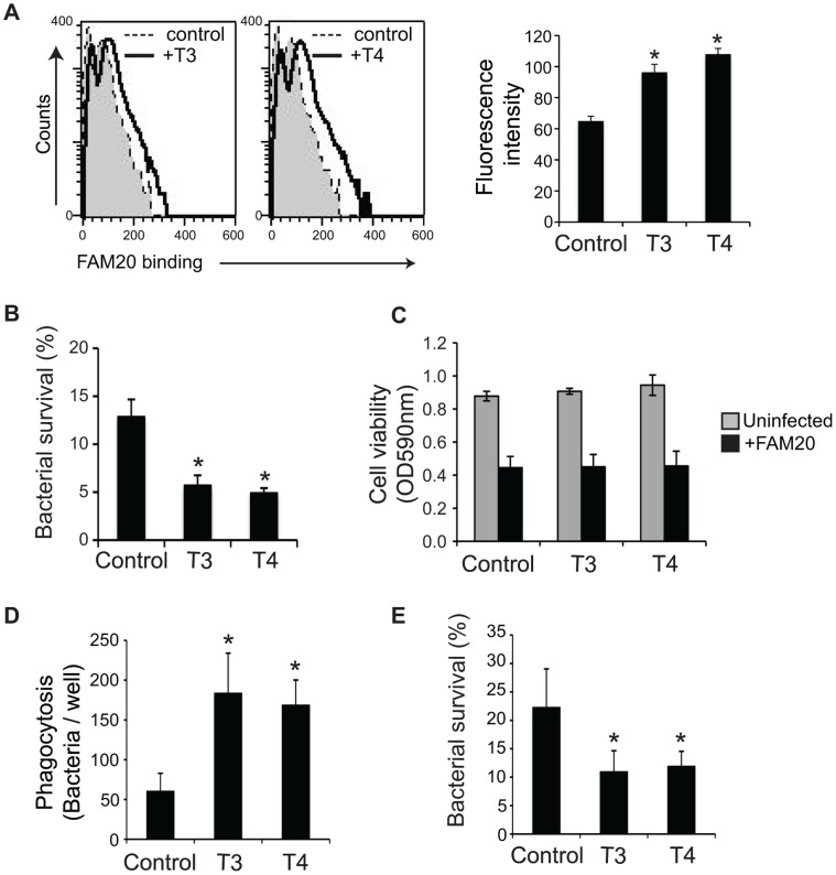
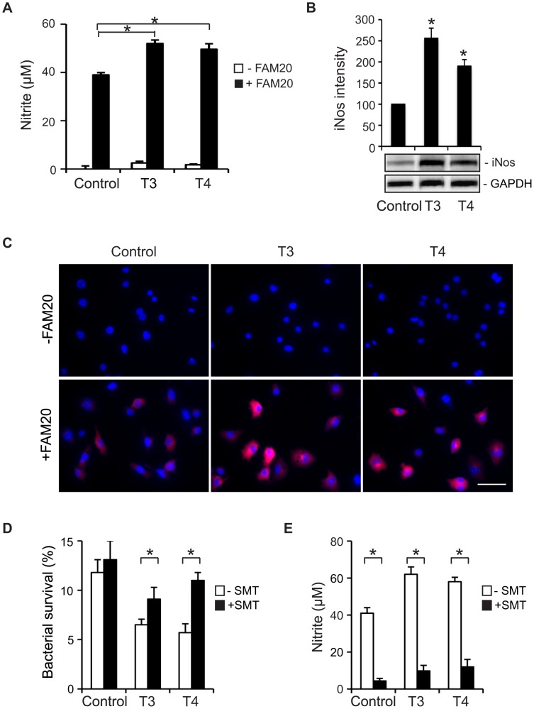
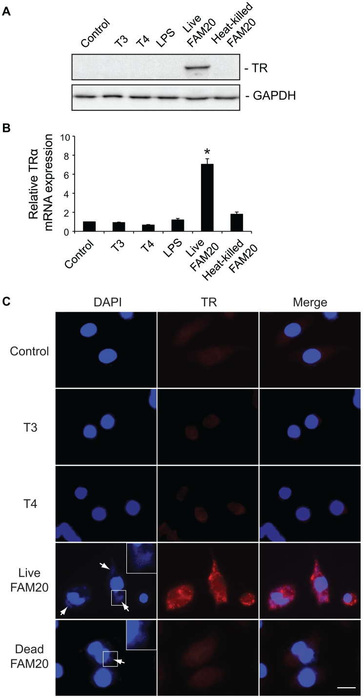
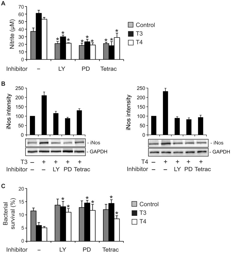
Euthyroid sick syndrome characterized by reduced levels of thyroid hormones (THs) is observed in patients with meningococcal shock. It has been found that the level of THs reflects disease severity and is predictive for mortality. The present study was conducted to investigate the impact of THs on host defense during meningococcal infection. We found that supplementation of thyroxine to mice infected with Neisseria meningitidis enhanced bacterial clearance, attenuated the inflammatory responses and promoted survival. In vitro studies with macrophages revealed that THs enhanced bacteria-cell interaction and intracellular killing of meningococci by stimulating inducible nitric oxide synthase (iNos)-mediated NO production. TH treatment did not activate expression of TH receptors in macrophages. Instead, the observed TH-directed actions were mediated through nongenomic pathways involving the protein kinases PI3K and ERK1/2 and initiated at the membrane receptor integrin αvβ3. Inhibition of nongenomic TH signaling prevented iNos induction, NO production and subsequent intracellular bacterial killing by macrophages. These data demonstrate a beneficial role of THs in macrophage-mediated N. meningitidis clearance. TH replacement might be a novel option to control meningococcal septicemia.


Conflict of interest statement

Competing Interests: The authors have declared that no competing interests exist.

Figures

Figure 1
Figure 1. T4 enhances survival during meningococcal infection.
(A-D) Mice (n = 10–12 per group) were treated with T4 (500 ng) or vehicle (control) for three days. On the second day of treatment, mice were challenged i.p. with 108 CFU of the N. meningitidis strain FAM20. (A) Survival of T4-treated and control mice after bacterial infection. Bacterial counts in blood (B), spleen (C) and liver (D) of mice at 24 h p.i. were determined. (E–F) Mice were challenged with 108 CFU of bacteria and treated with T4 (250–1000 ng/mouse) at 4 h and 24 h p.i. The control group was treated with vehicle. (E) Survival and (F) bacterial counts in blood of mice at 24 h p.i. were measured. *, P<0.05 (Nonparametric Mann-Whitney test in A and E, Student’s t-test in B–D, F). Symbols represent individual mice and the horizontal lines represent mean of the values.
Figure 2
Figure 2. T4 attenuates inflammatory responses in vivo.
Mice (n = 8 per group) were treated as described in Figure 1 and the concentrations of IL-6 (A), TNFα (B), KC (C), IFNγ (D), MIF (E) and C5a (F) in serum at 24 h p.i. were measured by ELISA. Symbols represent individual mice and the horizontal lines represent mean of the values. *, P<0.05 (Student’s t-test).
Figure 3
Figure 3. Impact of THs on macrophage cytokine production.
(A) Peritoneal macrophages were collected from mice treated with T4 or vehicle as described in Figure 1. (B) RAW264.7 cells and (C) PMA-differentiated THP-1 cells were treated with 100 nM T3 or 1 µM T4 for 24 h, cells treated with vehicle NaOH were set as control. Cells were infected with FAM20 at a MOI of 200 for 24 h and levels of IL-6 and TNFα in the supernatant were quantified by ELISA. The experiments were performed in triplicate and data are presented as means ±SD.
Figure 4
Figure 4. Impact of thyroid hormones on N. meningitidis growth.
(A) Effect of THs on meningococcal growth in GC liquid. FAM20 were suspended in GC liquid containing 100 nM T3 or 1 µM T4 and allowed to grow for up to 48 h. At indicated time points, the growth of bacteria was monitored by optical measurement at 600 nm. Bacteria grown in GC liquid were set as a control. (B) Effect of THs on meningococcal growth in serum. Serum was collected from mice treated with T4 or vehicle as described in Figure 1 and mixed with FAM20. As a control, DMEM was mixed with bacteria in the same way. The bacterial solutions were incubated for 1 h at 37°C, plated after serial dilutions and the number of surviving bacteria was counted the next day. Bacterial growth index was defined as (CFU after incubation)/(CFU before incubation) and bacterial growth in DMEM was set as 1. Experiments were performed in triplicate and data are presented as means ±SD.
Figure 5
Figure 5. TH enhances the bactericidal activity of macrophages.
(A)–(C) RAW264.7 cells were treated with 100 nM T3 or 1 µM T4 for 24 h. (A) Cells were infected with FAM20 (MOI  = 200) for 1 h at 4°C. Binding of the bacteria was determined by flow cytometry after staining with an anti-N. meningitidis Ab followed with an alexa 488-conjugated IgG. Mean fluorescence intensity of the entire population is presented. (B) TH-treated RAW264.7 cells were infected with FAM20 (MOI  = 200) for 3 h and intracellular bacterial survival was determined by a gentamicin protection assay as described in Materials and Methods. (C) Viability of TH-treated cells in the presence and absence of FAM20 was measured using a MTT assay. (D)-(E) PMA-differentiated THP-1 cells were treated with 100 nM T3 or 1 µM T4 for 24 h and then infected with FAM20 (MOI  = 200) for 3 h. (D) Phagocytosed bacteria and (E) Intracellular bacterial survival was determined by a gentamicin protection assay as described in Materials and Methods. Cells treated with vehicle NaOH were set as control. All experiments were performed in triplicate and data are presented as means ±SD. *, P<0.05 (Student’s t-test).
Figure 6
Figure 6. TH-enhanced bactericidal activity by macrophages is mediated through iNos/NO production.
(A)–(C) RAW264.7 cells were treated with 100 nM T3 or 1 µM T4 for 24 h prior to stimulation with heat-killed N. meningitidis FAM20 (MOI  = 200) for 24 h. Control cells were treated with vehicle for THs. (A) Production of NO was determined by measurement of nitrite in the cell supernatant. (B) Expression of iNos protein in cells was detected by Western blot using a rabbit anti-mouse iNos Ab. Expression of GAPDH was set as a loading control. Densitometric analysis of Western blot bands was performed using Image J software and relative expression levels of iNos protein was quantified after normalization to GAPDH. Data are presented as mean ±SD of three independent experiments and are indicated as percentage of control cells, which was set as 100%. *, P<0.05 (Student’s t-test). (C) Expression of iNos in cells was visualized by immunofluorescence microscopy using an anti-iNos Ab. Cell nuclei were stained with DAPI. Representative images from three independent experiments are shown. Scale bar: 40 µm. (D)–(E) RAW264.7 cells were cultured with 100 nM T3 or 1 µM T4 in the presence of (S)-methylisothiourea (SMT) for 24 h. Vehicle for THs (NaOH) or SMT (H2O) was added to control or SMT-untreated cells. (D) Cells were infected with live FAM20 (MOI  = 200) and intracellular bacterial survival was determined by a gentamicin protection assay as described in Materials and Methods. (E) Cells were infected with heat-killed FAM20 (MOI  = 200) for 24 h and nitrite in the cell supernatant was determined by a Griess assay. Experiments were performed in triplicate and data are presented as means ±SD. *, P<0.05 (Student’s t-test).
Figure 7
Figure 7. Expression of TRα in macrophages is induced by live N. meningitidis.
RAW264.7 cells were treated with T3 (100 nM), T4 (1 µM), LPS (500 ng/ml), live FAM20 (MOI  = 2) or heat-killed FAM20 (MOI  = 200) for 24 h. (A) Expression of TRα in cells was detected by Western blot using an anti-TRα/β Ab. Expression of GAPDH was measured as a loading control. (B) Levels of TRα mRNA were analyzed by real-time PCR. Expression levels were normalized to a reference mRNA (GAPDH). The experiment was performed in triplicate and results are presented as means ±SD. *, P<0.05 (Student’s t-test). (C) Expression and localization of TR in macrophages was visualized by immunofluorescence staining using an anti-TRα/β Ab. Cell nuclei and bacteria (arrows) were stained with DAPI. Representative images are shown from experiments that were repeated three times. Scale bar: 10 µm.
Figure 8
Figure 8. TR-independent pathways are involved in
TH-enhanced NO production and iNos expression. RAW264.7 cells were treated with or without (Control) 100 nM T3 or 1 µM T4 for 24 h in the presence or absence of 2.5 µM LY294002 (LY, PI3K inhibitor), 10 µM PD98059 (PD, ERK1/2 inhibitor) or 10 µM tetraiodothyroacetic acid (Tetrac, T3/T4 analog to integrin αvβ3 receptor). Vehicle for THs (NaOH) or inhibitors (DMSO) was added to control or inhibitor-untreated cells. (A) Cells were infected with heat-killed FAM20 (MOI  = 200) for 24 h. Levels of nitrite in the supernatants were determined by a Griess assay. *, P<0.05 (Student’s t-test, compared to cells without inhibitor treatment). (B) Cells were infected with heat-killed FAM20 (MOI  = 200) for 24 h. Expression of iNos protein in cells was detected by Western blot using an anti-iNos Ab. Densitometry of Western blot bands was performed using Image J software and relative expression level of iNos protein is presented after normalization to GAPDH. Data is displayed as arbitrary units set to 100 for the control without TH treatment. (C) RAW264.7 cells were treated as indicated above and infected with live FAM20 (MOI  = 200) for 3 h. Intracellular survival of meningococci was determined using a gentamicin protection assay as described in Materials and Methods. Experiments were performed in triplicate and data are presented as means ±SD. *, P<0.05 (Student’s t-test, compared to cells without inhibitor treatment).

References

    1. Kelly GS. Peripheral metabolism of thyroid hormones: a review. Altern Med Rev. 2000;5:306–333. - PubMed
    1. Friesema EC, Jansen J, Jachtenberg JW, Visser WE, Kester MH, et al. Effective cellular uptake and efflux of thyroid hormone by human monocarboxylate transporter 10. Mol Endocrinol. 2008;22:1357–1369. - PMC - PubMed
    1. Mitchell FE, Roy LA, Taylor PM. Iodothyronine Interactions with the System L1 Amino Acid Exchanger in 3T3-L1 Adipocytes. J Thyroid Res. 2010;2010:726098. - PMC - PubMed
    1. Flamant F, Gauthier K, Samarut J. Thyroid hormones signaling is getting more complex: STORMs are coming. Mol Endocrinol. 2007;21:321–333. - PubMed
    1. Bassett JH, Harvey CB, Williams GR. Mechanisms of thyroid hormone receptor-specific nuclear and extra nuclear actions. Mol Cell Endocrinol. 2003;213:1–11. - PubMed

Publication types

MeSH terms