. 2012;7(7):e41536.
doi: 10.1371/journal.pone.0041536. Epub 2012 Jul 23.

Protein tyrosine phosphatase PTP1B is involved in hippocampal synapse formation and learning

Affiliations

Protein tyrosine phosphatase PTP1B is involved in hippocampal synapse formation and learning

Federico Fuentes et al. PLoS One. 2012.

Abstract

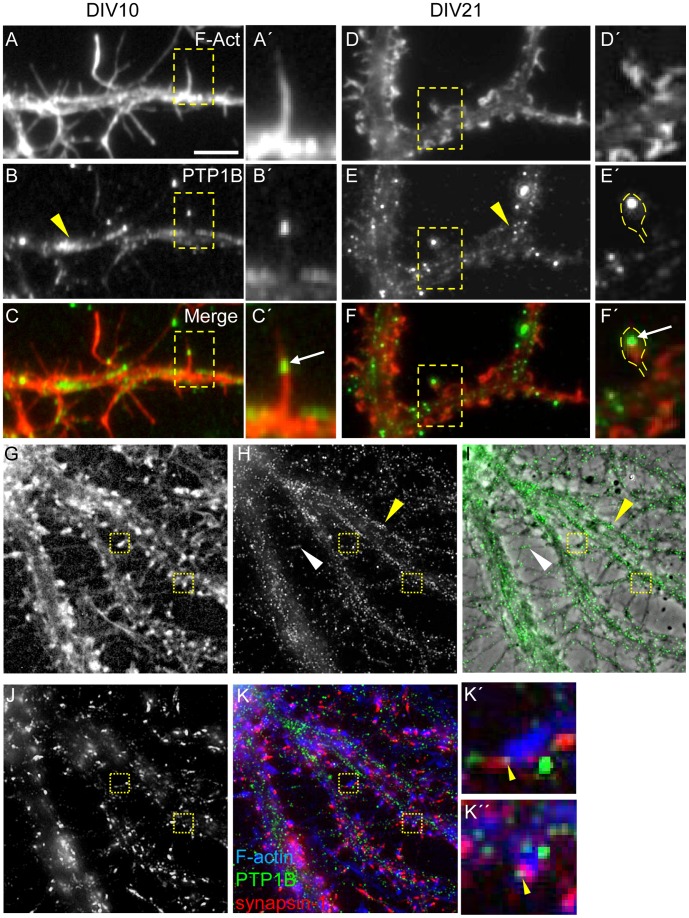
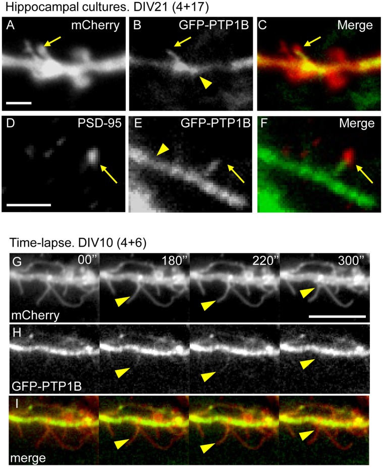
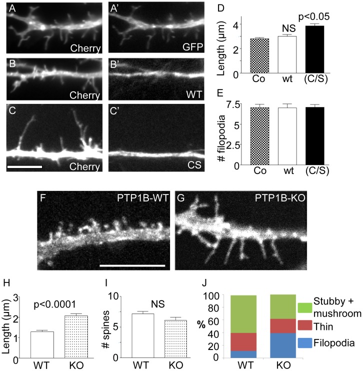
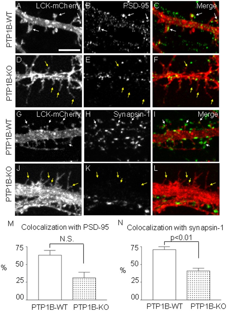
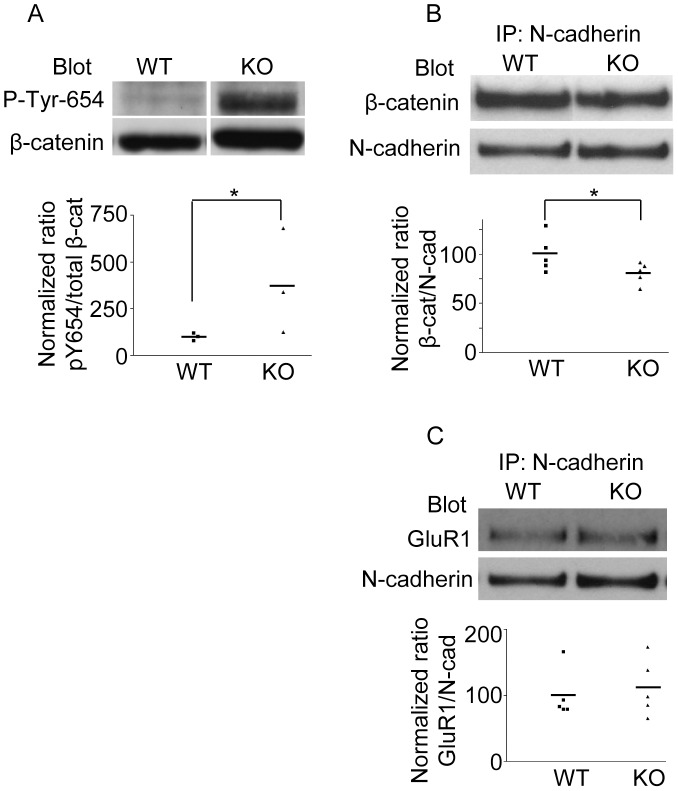
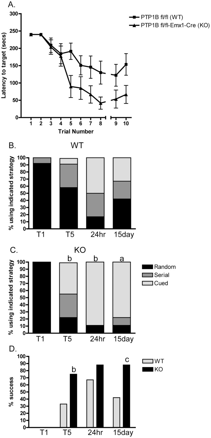
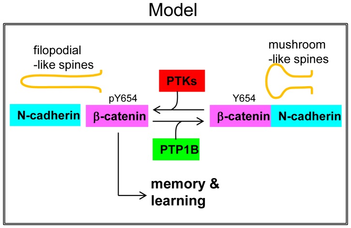
ER-bound PTP1B is expressed in hippocampal neurons, and accumulates among neurite contacts. PTP1B dephosphorylates ß-catenin in N-cadherin complexes ensuring cell-cell adhesion. Here we show that endogenous PTP1B, as well as expressed GFP-PTP1B, are present in dendritic spines of hippocampal neurons in culture. GFP-PTP1B overexpression does not affect filopodial density or length. In contrast, impairment of PTP1B function or genetic PTP1B-deficiency leads to increased filopodia-like dendritic spines and a reduction in mushroom-like spines, while spine density is unaffected. These morphological alterations are accompanied by a disorganization of pre- and post-synapses, as judged by decreased clustering of synapsin-1 and PSD-95, and suggest a dynamic synaptic phenotype. Notably, levels of ß-catenin-Tyr-654 phosphorylation increased ∼5-fold in the hippocampus of adult PTP1B(-/-) (KO) mice compared to wild type (WT) mice and this was accompanied by a reduction in the amount of ß-catenin associated with N-cadherin. To determine whether PTP1B-deficiency alters learning and memory, we generated mice lacking PTP1B in the hippocampus and cortex (PTP1B(fl/fl)-Emx1-Cre). PTP1B(fl/fl)-Emx1-Cre mice displayed improved performance in the Barnes maze (decreased time to find and enter target hole), utilized a more efficient strategy (cued), and had better recall compared to WT controls. Our results implicate PTP1B in structural plasticity within the hippocampus, likely through modulation of N-cadherin function by ensuring dephosphorylation of ß-catenin on Tyr-654. Disruption of hippocampal PTP1B function or expression leads to elongation of dendritic filopodia and improved learning and memory, demonstrating an exciting novel role for this phosphatase.


Conflict of interest statement

Competing Interests: The authors have declared that no competing interests exist.

Figures

Figure 1
Figure 1. Distribution of endogenous PTP1B during dendrite maturation.
Hippocampal neurons from rat embryos were cultured in serum-free medium and fixed at DIV10 (A–C) and DIV21 (D–K). Neurons were processed for fluorescence detection of F-actin, using phalloidin-TMR (A, A’, D, D’) or phalloidin-AMCA (G, K); PTP1B, using a specific mouse monoclonal antibody (B, B’, E, E’, H), and synapsin-1 using a rabbit polyclonal antibody (J). (A–C) At DIV10 most dendritic protrusions display a filopodial shape. (D–F) At DIV21 mushroom-shaped spines prevails. PTP1B displays a punctate distribution in dendritic shafts at both developmental stages (B, E, H, I, yellow arrowheads), and in thin axons (I, white arrowhead). Occasionally, PTP1B puncta locates in dendritic protrusions (white arrows in inset frames C’, F’). In neurons of DIV21, PTP1B puncta sometimes can be seen at the heads of mushroom-like spines (F’), co-localizing with synapsin-1 puncta (boxes, K’, K”, yellow arrowheads). Scale bar, 5 µm.
Figure 2
Figure 2. Localization and dynamics of GFP-PTP1B in dendritic protrusions.
Hippocampal neurons from rat embryos were co-transfected at DIV4 with plasmids encoding GFP-PTP1B and Lck-mCherry. (B, E, H) GFP-PTP1B signal is relatively strong and uniform in dendritic shafts of neurons imaged at DIV10 (H) and DIV21 (B, E, yellow arrowheads). In the DIV21 cultures, fingerlike protrusions of GFP-PTP1B emerge from dendrite shafts and penetrate into mushroom-like spines detected with the Lck-mCherry (A, B, C, yellow arrows). Scale bar, 2 µm in A. Note that the tips of the GFP-PTP1B protrusions co-localize with PSD-95 clusters detected by immunofluorescence (D, E, F, yellow arrows). Scale bar, 2 µm in D. (G–I) Time lapse studies in DIV10 cultures reveal a dynamic behavior of GFP-PTP1B, entering transiently to preformed dendritic filopodia (yellow arrowheads). Images were taken every 10 seconds during a 10 minute recording. Scale bar, 10 µm in G.
Figure 3
Figure 3. Effect of PTP1B inhibition on dendritic protrusions.
(A–E) Hippocampal neurons from rat embryos were co-transfected at DIV 4 with plasmids encoding Lck-mCherry and GFP, GFP-PTP1Bwt or the dominant negative GFP-PTP1B(C/S). At DIV 10 neurons were fixed and imaged for the fluorescent proteins. Expression of GFP-PTP1Bwt (wt) had no effect on filopodia length compared to GFP (B-B’ vs A–A’; scale bar, 5 µm in C). In contrast, expression of GFP-PTP1B(C/S) leads to a significant increase of filopodia length (C–C’). (D) Plot showing the quantification of filopodia length. (E) Plot showing the density of filopodia per 10 µm. (F–J) Hippocampal neurons from PTP1B KO and wild type (WT) newborn mice were transfected at DIV4 with a plasmid encoding Lck-mCherry. At DIV14, neurons were fixed and observed in a fluorescence microscope. Note the predominance of filopodia-like protrusions in the KO neurons (G) compared to WT neurons (F; Scale bar, 5 µm). (H) Quantification of the length of dendritic protrusions shows a significant increase in KO neurons compared to WT neurons. (I) Density of spines does not differ significantly. (J) Quantification of the different morphological types of spines reveals that KO neurons had a significantly reduced proportion of stubby and mushroom spines, and a significantly increased proportion of filopodia-like protrusions, compared to WT neurons. ANOVA p<0.0001 followed by a Dunnett’s post-test p<0.05.
Figure 4
Figure 4. Effect of PTP1B deficiency on the distribution of pre-and post-synaptic markers.
Hippocampal neurons from WT (A–C, G–I) and KO (D–F, J–L) newborn mice were transfected at DIV4 with Lck-mCherry to visualize dendritic spines. At DIV14 neurons were fixed and immunostained to detect either the post-synaptic marker PSD-95 (B, E) or the pre-synaptic marker synapsin-1 (H, K). PSD-95 and synapsin-1 were visualized using Alexa Fluor 488-conjugated secondary antibodies. Localization of PSD-95 in spine heads is obvious in WT neurons (A–C, white arrows). In contrast, filopodia-like protrusions from KO neurons show no PSD-95 associated (D–F, yellow arrows). Instead, PSD-95 clusters were found in dendritic shafts. Synapsin-1 clusters are also evident adjacent to spines heads in WT neurons (G–I, white arrows) but not to filopodia-like protrusions in KO neurons (J–L, yellow arrows). Scale bar, 5 µm in A. Plots show the quantification of dendritic protrusions showing colocalization with (M) PSD-95 (WT 63±6.6% versus KO 31±7.7%, p = 0.06) and (N) synapsin-1 puncta (WT 70.8±4.2% versus KO 41.1±3.5%, p = 0.01).
Figure 5
Figure 5. PTP1B controls ß-catenin phosphorylation and association with N-cadherin in vivo.
Protein extracts from hippocampi of adult WT and KO mice were prepared. (A) Western blots were first probed with a polyclonal antibody specific for ß-catenin-pTyr-654. Subsequently, the membrane was stripped and re-probed with a monoclonal antibody against total ß-catenin. The normalized pY654/ß-catenin signal was calculated from scanned bands. Note that KO mice show a significant increase of the normalized signal compared to the WT mice (KO: 380.5±161.7% vs WT: 100.0±11.6%). (B) N-cadherin was immunoprecipitated using a specific monoclonal antibody. Western blots of N-cadherin immunoprecipitates were first probed to detect total ß-catenin and then re-probed to detect N-cadherin. Experiments from five animals show that normalized ratios of ß-catenin/N-cadherin in KO mice are significantly reduced compared to those in WT mice (KO: 80.8±7% vs WT: 100±3, p<0.05 one-tailed Mann-Whitney test). (C) Amount of GluR1 co-immunoprecipitated with N-cadherin. Experiments from five animals show that normalized ratios of GluR1/N-cadherin are not statistically different between WT and KO mice (KO: 112.1±19.2% vs WT: 100.0±16.8%. Asterisks indicate statistical differences for a p≤0.05, according to the one-tailed Mann-Whitney test.
Figure 6
Figure 6. PTP1B fl/fl-Emx1-Cre and littermate PTP1B fl/fl controls were subjected to the Barnes maze.
(A) Mice were trained on the maze 2 trials per day for 4 days and their performance plotted as time to enter the target escape hole (trials 1–8). A 24 hour recall (trial 9) and a 15 day recall (trial 10) were performed to assess memory retention. n = 12 WT and n = 8 KO. Data are mean ± SEM, asterisk indicates p<0.01 by 2-way ANOVA. (B and C) Female PTP1B fl/fl (WT; n = 12) and PTP1B fl/fl-Emx1-Cre (KO; n = 8) littermate controls were scored for strategy used in the Barnes maze and plotted as the percentage of mice using a random, serial, or cued strategy to locate the target hole. (D) The percentage of mice successfully locating the target hole in each trial/recall is plotted. T1, trial 1; T5, trial 5; 24 hr, 24 hour recall; 15 day, 15 day recall. a, p<0.05; b, p<0.08; c, p = 0.06 by a nominal logistic followed by a Pearson’s Chi-square test comparing WT and KO for the indicated trial.
Figure 7
Figure 7. Model representing the potential function of PTP1B in the hippocampus.
PTP1B dephosphorylates ß-catenin at the residue Tyr-654, opposing the activity of protein tyrosine kinases. This function of PTP1B ensures ß-catenin association with N-cadherin in functional adhesion complexes, and is required for normal differentiation of mushroom-like spines. Loss of PTP1B expression/function would favor a stage characterized by morphologically immature (and likely more dynamic) spines, which may positively modulate memory and learning processes.

References

    1. Lamprecht R, LeDoux J. Structural plasticity and memory. Nat Rev Neurosci. 2004;5:45–54. - PubMed
    1. Yuste R, Bonhoeffer T. Genesis of dendritic spines: insights from ultrastructural and imaging studies. Nat Rev Neurosci. 2004;5:24–34. - PubMed
    1. Bourne JN, Harris KM. Balancing structure and function at hippocampal dendritic spines. Annu Rev Neurosci. 2008;31:47–67. - PMC - PubMed
    1. Holtmaat A, Svoboda K. Experience-dependent structural synaptic plasticity in the mammalian brain. Nat Rev Neurosci. 2009;10:647–658. - PubMed
    1. Leuner B, Gould E. Structural plasticity and hippocampal function. Annu Rev Psychol. 2010;61:111–140. - PMC - PubMed

Publication types

MeSH terms