. 2012;7(7):e41688.
doi: 10.1371/journal.pone.0041688. Epub 2012 Jul 26.

MicroRNA regulation of the synaptic plasticity-related gene Arc

Affiliations

MicroRNA regulation of the synaptic plasticity-related gene Arc

Karin Wibrand et al. PLoS One. 2012.

Abstract

Expression of activity-regulated cytoskeleton associated protein (Arc) is crucial for diverse types of experience-dependent synaptic plasticity and long-term memory in mammals. However, the mechanisms governing Arc-specific translation are little understood. Here, we asked whether Arc translation is regulated by microRNAs. Bioinformatic analysis predicted numerous candidate miRNA binding sites within the Arc 3'-untranslated region (UTR). Transfection of the corresponding microRNAs in human embryonic kidney cells inhibited expression of an Arc 3'UTR luciferase reporter from between 10 to 70% across 16 microRNAs tested. Point mutation and deletion of the microRNA-binding seed-region for miR-34a, miR-326, and miR-19a partially or fully rescued reporter expression. In addition, expression of specific microRNA pairs synergistically modulated Arc reporter expression. In primary rat hippocampal neuronal cultures, ectopic expression of miR-34a, miR-193a, or miR-326, downregulated endogenous Arc protein expression in response to BDNF treatment. Conversely, treatment of neurons with cell-penetrating, peptide nucleic acid (PNA) inhibitors of miR-326 enhanced Arc mRNA expression. BDNF dramatically upregulated neuronal expression of Arc mRNA and miR-132, a known BDNF-induced miRNA, without affecting expression of Arc-targeting miRNAs. Developmentally, miR-132 was upregulated at day 10 in vitro whereas Arc-targeting miRNAs were downregulated. In the adult brain, LTP induction in the dentate gyrus triggered massive upregulation of Arc and upregulation of miR-132 without affecting levels of mature Arc-targeting miRNAs. Turning to examine miRNA localization, qPCR analysis of dentate gyrus synaptoneurosome and total lysates fractions demonstrated synaptic enrichment relative to small nucleolar RNA. In conclusion, we find that Arc is regulated by multiple miRNAs and modulated by specific miRNA pairs in vitro. Furthermore, we show that, in contrast to miR-132, steady state levels of Arc-targeting miRNAs do not change in response to activity-dependent expression of Arc in hippocampal neurons in vitro or during LTP in vivo.


Conflict of interest statement

Competing Interests: The authors have declared that no competing interests exist.

Figures

Figure 1
Figure 1. Luciferase reporter screen for microRNAs targeting the Arc 3′UTR.
A) Arc 3′UTR luciferase reporter vector and microRNA precursor plasmids were co-transfected in HEK293T cells. After 48 h the luciferase activity was measured and normalized to transfection control (gWIZ, alkaline phosphatase). The luciferase values were further normalized to the average luciferase value obtained after transfecting a panel of microRNAs not predicted to target the rat Arc 3′UTR (rno-miR-370, rno-miR-150, rno-miR-342, rno-miR-30b, rno-miR-105, rno-miR-145 and rno-miR-9). The candidate Arc-targeting microRNAs produced a graded inhibition of Arc 3′UTR luciferase expression ranging from 0 to 70%. B) Schematic representation of the localization of the seven predicted microRNA binding sites in the Arc 3′UTR (NM_019361) that were selected for further studies with site-directed mutagenesis. The numbering refers to the position in the 3′UTR. The nucleotides that were changed by site-directed mutagenesis are underlined in the alignment.
Figure 2
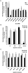
Figure 2. The effects of miR-19a and miR-34a and miR-326 is dependent on intact microRNA binding sites.
A) Site-directed mutagenesis was carried out to interfere with 6 different microRNA sites in the Arc 3′UTR. Three nucleotides in the seed-binding region of miR-34, -193, -326, -378 and -512_5p were mutated in the Arc 3′UTR and the whole seed binding region was removed for miR-19. 48 h after co-transfection of HEK293T cells with Arc reporter constructs (wildtype or mutated) and microRNA expression vectors, the medium was harvested for measurement of luciferase and SEAP activity. A significant difference in luciferase expression was observed after substitution mutation or deletion of the miRNA binding sites for miR-19a, miR-34a and miR-326 in response to expression of the respective miRNAs, relative to the wildtype Arc 3′UTR. B) Deletion of the miR-34a site resulted in full recovery of luciferase activity. The positive control comprised of a fully complementary sensor sequence of miR-34a was efficiently inhibited by miR-34a overexpression. C) Whereas mutation of the distal (S4) miR-326 site or deletion of either site individually had only minor effects, the deletion of both sites at the same time gave full recovery of luciferase activity. The sensor construct of miR-326 showed the same effect as deleting both miR-326 binding sites. In panels A, B and C luciferase expression was normalized to the transfection control (gWIZ, alkaline phosphatase) and to miR-150. miR-150 was one of the miRNAs in the initial screen with least effect on the luciferase activity of the reporter vector. Values are means ± SEM (n = 3). * p<0.05, significantly different from wildtype (Student's t-test).
Figure 3
Figure 3. Repression of Arc is enhanced by expression of microRNA pairs.
Arc-targeting microRNAs were overexpressed in HEK293T cells alone or in pairs. The following combinations: miR-34a/miR-193a, miR-326/378 and miR-326/193a gave enhanced inhibition of luciferase activity compared to miR-34a and miR-326 alone. Values are means ± SEM (n = 3). *p<0.05, significantly different from miR-34a or miR-326 alone (Student's t-test).
Figure 4
Figure 4. miR-34a, miR-326 and miR-193a downregulate Arc protein expression in cultured hippocampal neurons.
Cultured hippocampal neurons were transfected with either empty vector-DsRed, miR150-DsRed, miR34a-DsRed, miR326-DsRed or miR193a-DsRed. Neurons were treated with BDNF for four hours and Arc protein expression was assessed by light microscopy. A) representative images of cells transfected with empty vector-DsRed and miR34a-DsRed, respectively. Note that Arc levels are very diverse within one sample. The fields of view were chosen to contain at least one DsRed-expressing neuron per image, and approximately 60 images from at least two different coverslips were taken per condition per experiment. During image acquisition and subsequent data analysis, the experimenter was blinded to the treatment group of the cells. B) scatter plots of Arc vs. DsRed levels per cell. Data are normalized to the average value of non-transfected cells. Gray diamonds = non-transfected cells; black diamonds = transfected cells. n = 3996 cells from 3 experiments. C) bar graph comparing the average Arc level in transfected cells. Values are normalized to the average Arc level in the non-transfected cells. A significant downregulation of Arc protein was seen in miR-34a transfected cells. Significance was tested by independent t-tests, p<0.001. Error bars = SEM. n = 763 cells from 3 experiments. D) representative images of cells transfected with miR-326-DsRed (cells labelled with DsRed and red arrows) and non-transfected cells (white arrows). Immunostaining of Arc protein is visualized in blue. Note that Arc levels are very diverse within one sample. Phalloidin labelling in green was used to visualize the individual cells. E) bar graph comparing the average Arc level in transfected cells. Values are normalized to the average Arc level in the non-transfected cells. A significant downregulation of Arc protein was seen in miR-193a and miR-326 transfected cells. Significance was tested by univariate ANOVA and post hoc tests, p<0.05. Error bars = SEM. n = 339 cells from 3 experiments.
Figure 5
Figure 5. Inhibition of endogenous Arc-targeting miRNAs in hippocampal neurons.
PNA-conjugated antisense oligonucleotides were used to block endogenous microRNAs. A) The uptake of a fluorescent control PNA oligo is efficient in hippocampal neurons (DIV8) 24 hours after transfection. Green fluorescence = PNA. DsRed was cotransfected to compare the level of PNA transfection with that of plasmid. B) RNA enriched for small RNAs was isolated 48 h after transfection of antisense and scrambled control PNA and the level of unbound microRNAs was assayed by real-time PCR. 1% of miR-326, 0.7% of miR-34a and 4.7% of miR-193a remained unblocked after specific PNA transfection compared to control. Y1 was used for normalization. Values are means of n = 3–4 ± SEM C) RNA was isolated 48 h after transfection of microRNA inhibitors and used for Arc mRNA Q-PCR. Arc mRNA is significantly higher after transfection of miR-326 PNA-AS compared to scrambled (p = 0.04). Significance was tested by independent t-tests. Data is normalized to polyubiquitin and cyclophilin. Values are means of n = 5 ± SEM. D) Proteins were harvested 48 h after PNA transfection. Bar graphs representing densitometry measurements from protein western blot analysis. The Arc expression was normalized to the expression of GAPDH. Values are means of n = 7 ± SEM.
Figure 6
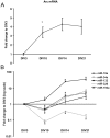
Figure 6. Developmental regulation of Arc-targeting microRNAs.
Arc mRNA expression and mature microRNA levels in hippocampal neurons from E18 rat embryos at different developmental stages (days in vitro, DIV). A) Quantitative relative real-time PCR of Arc mRNA. Arc mRNA increased at early time points and reached a plateau around DIV14. The relative values are expressed as fold change to DIV3 and normalized to the geometric mean of the reference genes HRPT and cyclophilin. B) Quantitative relative real-time PCR of miR-19a, miR-34a, miR-326, miR-193a and miR-132. The relative values are expressed as fold change to DIV3 and normalized to the reference genes snoRNA and Y1. At early stages of differentiation there was an inverse relation between the expression of Arc mRNA and miR-34a, -19a, -326 and -193a expression. miR-132 expression increases with neuronal differentiation. Note that the y-axis scale is in log format in B. In both A and B significance was tested by independent t-tests, p<0.05. Values are means of n = 4 ± SEM. * significantly different from the preceding time point.
Figure 7
Figure 7. BDNF does not alter levels of mature Arc-targeting miRNAs.
The effect of BDNF stimulation (for 30 minutes or 3 hours) on microRNA and Arc expression was studied at DIV8. A) Quantitative relative real-time PCR of Arc mRNA. The relative values are expressed as fold change to untreated control cells and normalized to the reference genes HRPT, polyubiquitin and cyclophilin. Arc mRNA levels are significantly increased at 30 minutes and 2 hours of BDNF treatment. B) Quantitative relative real-time PCR of miR- miR-133, 19a, miR-34a, miR-326 and miR-193a. The relative values are expressed as fold change to untreated control cells and normalized to the reference genes snoRNA and Y1. The expression of Arc-targeting miRNAs did not change in response to BDNF. Values are means of n = 4 ± SEM. In both A and B significance was tested by independent t-tests, p<0.05. * significantly different from control.
Figure 8
Figure 8. LTP-inducing stimulation does not alter the levels of mature Arc-targeting miRNAs.
The effect of LTP induction in the dentate gyrus on miRNA and Arc expression was studied by real-time PCR analysis of dentate gyrus samples obtained 30 min and 3 hours after high-frequency stimulation (HFS) of the perforant path. A) Real-time PCR of Arc mRNA. Bar graphs indicate fold change in the HFS-treated dentate gyrus relative to the control, contralateral dentate gyrus. Arc mRNA is induced 80-fold after HFS. Values are means of n = 2 ± SEM. The data was normalized to the reference genes HRPT, polyubiquitin and cyclophilin. B) Quantitative relative real-time PCR of miR-19a, miR-34a, miR-326, miR-193a and miR-132. miR-132 was significantly elevated at 2 h post-HFS. Bar graphs indicate fold change values comparing treated dentate gyrus relative to control. Values are means of n = 5 ± SEM. Data has been normalized to the reference genes Y1. Significance was tested by independent t-tests. * p<0.05 and significantly different from control.
Figure 9
Figure 9. Synaptic expression of Arc-targeting microRNAs.
A) Synaptoneurosomes (SNs) were prepared from normal untreated dentate gyrus and semi- quantitative relative real-time PCR was used to assess the synaptic localization of Arc-targeting miRNAs. Selected miRNAs were assayed in total homogenate and in the synaptoneurosome fraction. miR-124a was equally expressed in both preparations and used to normalize the data. Small nucleolar RNA (snoRNA) was highly depleted from the SN fraction and served as a quality control of the preparation. In contrast, the Arc-associated miRNAs and a known dendritic miRNA, miR-132, showed similar SN/homogenate expression ratios. The relative values are expressed as the fold enrichment between synaptoneurosomes and total homogenate. n = 6 Significance was tested by independent t-tests, p<0.05. *Significantly different from total homogenate. Error bars = SEM. B) miRNA in situ hybridization was performed on coronal hippocampal sections using LNA probes for miR-326, miR-34a, and scrambled control. miR-326 and miR-34a showed specific staining in the cell body layers of the dentate gyrus and cornu ammonis (CA) regions compared to scramble control. For miR-34a, the staining was observed approximately 30 µm into the apical dendrites of CA1 pyramidal cells. Lower right panel shows CA1 region with examples of proximal dendritic staining marked by white arrows.

Similar articles

Cited by

References

    1. Bramham CR, Worley PF, Moore MJ, Guzowski JF. The immediate early gene arc/arg3.1: regulation, mechanisms, and function. J Neurosci. 2008;28((46)):11760–11767. - PMC - PubMed
    1. Korb E, Finkbeiner S. Arc in synaptic plasticity: from gene to behavior. Trends Neurosci. 2011;34:591–598. - PMC - PubMed
    1. Bramham CR, Alme MN, Bittins M, Kuipers SD, Nair RR, et al. The Arc of synaptic memory. Exp Brain Res. 2010;200:125–140. - PMC - PubMed
    1. Shepherd JD, Bear MF. New views of Arc, a master regulator of synaptic plasticity. Nat Neurosci. 2011;14:279–284. - PMC - PubMed
    1. Plath N, Ohana O, Dammermann B, Errington ML, Schmitz D, et al. Arc/Arg3.1 is essential for the consolidation of synaptic plasticity and memories. Neuron. 2006;52((3)):437–444. - PubMed

Publication types

MeSH terms

Substances