

Overexpression of the DEC1 protein induces senescence in vitro and is related to better survival in esophageal squamous cell carcinoma
- PMID: 22844531
- PMCID: PMC3402465
- DOI: 10.1371/journal.pone.0041862
Overexpression of the DEC1 protein induces senescence in vitro and is related to better survival in esophageal squamous cell carcinoma
Abstract
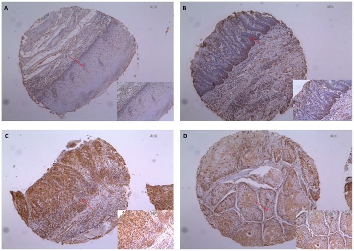
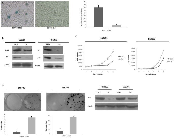
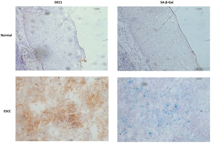
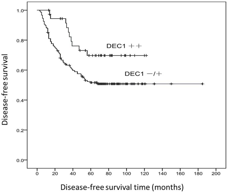
Esophageal squamous cell carcinoma (ESCC) is a leading cause of cancer-related death in China and has limited effective therapeutic options except for early surgery, since the underlying molecular mechanism driving its precursor lesions towards invasive ESCC is not fully understood. Cellular senescence is the state of the permanent growth arrest of a cell, and is considered as the initial barrier of tumor development. Human differentiated embryo chondrocyte expressed gene 1 (Dec1) is an important transcription factor that related to senescence. In this study, DEC1 immunohistochemical analysis was performed on tissue microarray blocks constructed from ESCC combined with adjacent precursor tissues of 241 patients. Compared with normal epithelia, DEC1 expression was significantly increased in intraepithelial neoplasia and DEC1 expression was significantly decreased in ESCC in comparison with intraepithelial neoplasia. In vitro, DEC1 overexpression induced cellular senescence, and it inhibited cell growth and colony formation in ESCC cell line EC9706. Fresh esophagectomy tissue sections from five ESCC patients were detected by immunohistochemistry of DEC1 and senescence-associated β-galactosidase (SA-β-Gal) activity, and strongly positive expression of DEC1 was correlated to more senescent cells in these fresh tissue sections. Kaplan-Meier method analysis of the 241 patients revealed that DEC1 expression levels were significantly correlated with the survival of ESCC patients after surgery. The expression levels of DEC1 were also correlated with age, tumor embolus, depth of invasion of ESCC, lymph metastasis status and pTNMs. These results suggest that DEC1 overexpression in precursor lesions of ESCC is a protective mechanism by inducing cellular senescence in ESCC initiation, and DEC1 may be a potential prognostic marker of ESCC.
Conflict of interest statement
Figures
Similar articles
-
Frequent decreased expression of candidate tumor suppressor gene, DEC1, and its anchorage-independent growth properties and impact on global gene expression in esophageal carcinoma.Int J Cancer. 2008 Feb 1;122(3):587-94. doi: 10.1002/ijc.23144. Int J Cancer. 2008. PMID: 17943723
-
Abnormal Localization and Tumor Suppressor Function of Epithelial Tissue-Specific Transcription Factor ESE3 in Esophageal Squamous Cell Carcinoma.PLoS One. 2015 May 7;10(5):e0126319. doi: 10.1371/journal.pone.0126319. eCollection 2015. PLoS One. 2015. PMID: 25950810 Free PMC article.
-
RNF113A promotes the proliferation, migration and invasion, and is associated with a poor prognosis of esophageal squamous cell carcinoma.Int J Oncol. 2018 Mar;52(3):861-871. doi: 10.3892/ijo.2018.4253. Epub 2018 Jan 24. Int J Oncol. 2018. PMID: 29393393
-
Upregulation of the long non-coding RNA BANCR correlates with tumor progression and poor prognosis in esophageal squamous cell carcinoma.Biomed Pharmacother. 2016 Aug;82:406-12. doi: 10.1016/j.biopha.2016.05.014. Epub 2016 May 31. Biomed Pharmacother. 2016. PMID: 27470379
-
Therapy-induced senescence as a component of tumor biology: Evidence from clinical cancer.Biochim Biophys Acta Rev Cancer. 2023 Nov;1878(6):188994. doi: 10.1016/j.bbcan.2023.188994. Epub 2023 Oct 6. Biochim Biophys Acta Rev Cancer. 2023. PMID: 37806641 Review.
Cited by
-
Cellular senescence in cancer: clinical detection and prognostic implications.J Exp Clin Cancer Res. 2022 Dec 27;41(1):360. doi: 10.1186/s13046-022-02555-3. J Exp Clin Cancer Res. 2022. PMID: 36575462 Free PMC article. Review.
-
Regulation of the Mechanism of TWIST1 Transcription by BHLHE40 and BHLHE41 in Cancer Cells.Mol Cell Biol. 2015 Dec;35(24):4096-109. doi: 10.1128/MCB.00678-15. Epub 2015 Sep 21. Mol Cell Biol. 2015. PMID: 26391953 Free PMC article.
-
Differentiated Embryo-Chondrocyte Expressed Gene1 and Parkinson's Disease: New Insights and Therapeutic Perspectives.Curr Neuropharmacol. 2023;21(11):2251-2265. doi: 10.2174/1570159X21666230502123729. Curr Neuropharmacol. 2023. PMID: 37132111 Free PMC article. Review.
-
Werner syndrome through the lens of tissue and tumour genomics.Sci Rep. 2016 Aug 25;6:32038. doi: 10.1038/srep32038. Sci Rep. 2016. PMID: 27559010 Free PMC article.
-
The transcription factor DEC1 (BHLHE40/STRA13/SHARP-2) is negatively associated with TNM stage in non-small-cell lung cancer and inhibits the proliferation through cyclin D1 in A549 and BE1 cells.Tumour Biol. 2013 Jun;34(3):1641-50. doi: 10.1007/s13277-013-0697-z. Epub 2013 Feb 20. Tumour Biol. 2013. PMID: 23423709
References
-
- Ferlay J, Shin HR, Bray F, Forman D, Mathers C, et al. Estimates of worldwide burden of cancer in 2008: GLOBOCAN 2008. Int J Cancer. 2010;127:2893–2917. - PubMed
-
- Jemal A, Bray F Center MM, Ferlay J, Ward E, et al. Global cancer statistics. CA Cancer J Clin. 2011;61:69–90. - PubMed
-
- Shirakawa Y, Naomoto Y, Kimura M, Kawashima R, Yamatsuji T, et al. Topological analysis of p21WAF1/CIP1 expression in esophageal squamous dysplasia. Clin Cancer Res. 2000;6:541–550. - PubMed
Publication types
MeSH terms
Substances
LinkOut - more resources
Full Text Sources
Medical
