

Cutaneous T-cell lymphoma in asians
- PMID: 22844610
- PMCID: PMC3403505
- DOI: 10.5402/2012/575120
Cutaneous T-cell lymphoma in asians
Abstract
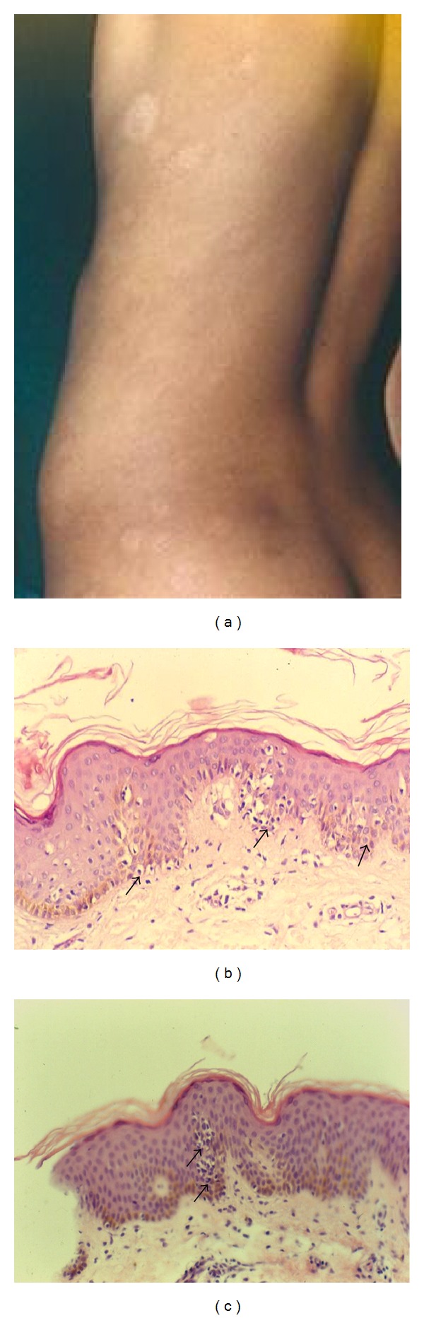
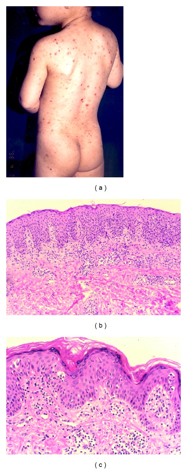
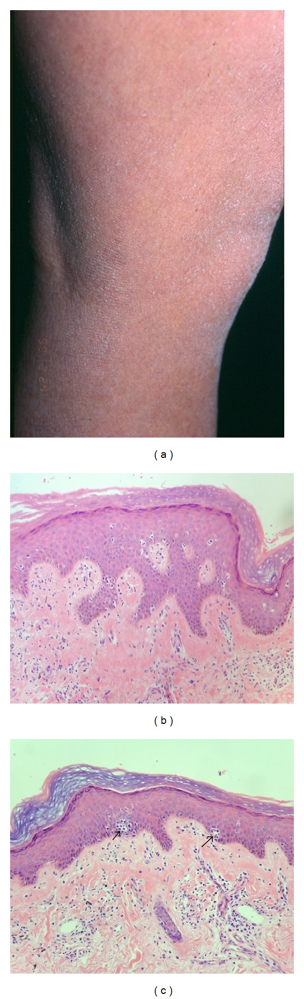
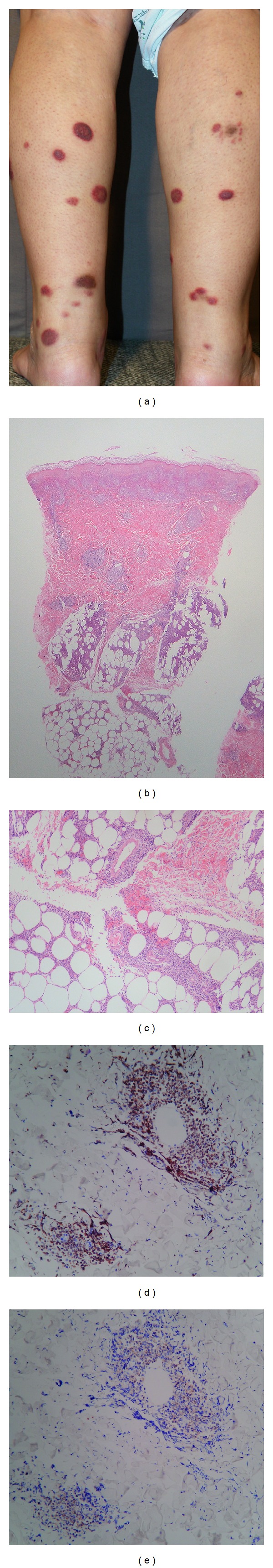
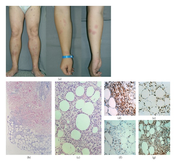
Cutaneous T-cell lymphoma describes a heterogeneous group of neoplasms of skin homing T cells that vary considerably in clinical presentation, histologic appearance, immunophenotype, and prognosis. This paper addresses the cutaneous T-cell lymphoma in Asians with respect to clinical-epidemiologic and histopathological features. Compared with Western countries, Asia usually has higher rates of cutaneous T-cell lymphomas such as extranodal NK/T-cell lymphoma, hydroa vacciniforme-like lymphoma, subcutaneous panniculitis T-cell lymphoma, and adult T-cell leukemia/lymphoma and lower rates of cutaneous B-cell lymphomas. Among many variants of mycosis fungoides, hypopigmented lesions, pityriasis lichenoides-like lesions, and ichthyosiform lesions are more prevalent in Asia than in the West. Adult T-cell leukemia/lymphoma is endemic in southwestern Japan especially in the Kyushu island. The clinicopathologic characteristics of cutaneous lymphoma vary according to geography, and this may be ascribed to genetic and environmental etiologic factors.
Figures
Similar articles
-
Primary cutaneous T-cell lymphoma: experience from the Peruvian National Cancer Institute.An Bras Dermatol. 2017 Sep-Oct;92(5):649-654. doi: 10.1590/abd1806-4841.20176825. An Bras Dermatol. 2017. PMID: 29166501 Free PMC article.
-
The spectrum of cutaneous lymphomas in patients less than 20 years of age.Pediatr Dermatol. 2004 Sep-Oct;21(5):525-33. doi: 10.1111/j.0736-8046.2004.21500.x. Pediatr Dermatol. 2004. PMID: 15461755
-
Non-mycosis fungoides cutaneous T-cell lymphomas: report of the 2011 Society for Hematopathology/European Association for Haematopathology workshop.Am J Clin Pathol. 2013 Apr;139(4):491-514. doi: 10.1309/AJCP83AOQTMLOJTM. Am J Clin Pathol. 2013. PMID: 23525618
-
Subcutaneous, blastic natural killer (NK), NK/T-cell, and other cytotoxic lymphomas of the skin: a morphologic, immunophenotypic, and molecular study of 50 patients.Am J Surg Pathol. 2004 Jun;28(6):719-35. doi: 10.1097/01.pas.0000126719.71954.4f. Am J Surg Pathol. 2004. PMID: 15166664
-
Cutaneous T-cell lymphomas (including rare subtypes). Current concepts. II.Haematologica. 2004 Nov;89(11):1372-88. Haematologica. 2004. PMID: 15531460 Review.
Cited by
-
Characteristics of Primary Cutaneous T-Cell Lymphoma in Iran: A 10-Year Retrospective Study.Int Sch Res Notices. 2014 Dec 24;2014:820921. doi: 10.1155/2014/820921. eCollection 2014. Int Sch Res Notices. 2014. PMID: 27437467 Free PMC article.
-
Subcutaneous Panniculitis-like T-Cell Lymphoma with a Transformation to Lupus Erythematosus Panniculitis: A Case Report.Case Rep Dermatol. 2022 Nov 4;14(3):319-325. doi: 10.1159/000527530. eCollection 2022 Sep-Dec. Case Rep Dermatol. 2022. PMID: 36466756 Free PMC article.
-
Hydroa Vacciniforme and Hydroa Vacciniforme-Like Lymphoproliferative Disorder: A Spectrum of Disease Phenotypes Associated with Ultraviolet Irradiation and Chronic Epstein-Barr Virus Infection.Int J Mol Sci. 2020 Dec 7;21(23):9314. doi: 10.3390/ijms21239314. Int J Mol Sci. 2020. PMID: 33297336 Free PMC article. Review.
-
Systemic lymphoma arising from hydroa vacciniforme-like lymphoma: report of two cases with review of literature.Int J Clin Exp Pathol. 2014 Aug 15;7(9):6403-8. eCollection 2014. Int J Clin Exp Pathol. 2014. PMID: 25337300 Free PMC article. Review.
-
Defining the mimics and clinico-histological diagnosis criteria for mycosis fungoides to minimize misdiagnosis.Int J Womens Dermatol. 2017 Jan 30;3(2):100-106. doi: 10.1016/j.ijwd.2016.11.006. eCollection 2017 Jun. Int J Womens Dermatol. 2017. PMID: 28560304 Free PMC article.
References
-
- Criscione VD, Weinstock MA. Incidence of cutaneous T-cell lymphoma in the United States, 1973–2002. Archives of Dermatology. 2007;143(7):854–859. - PubMed
-
- Lee MW, Korean Dermatopathology Research Group Characteristics of cutaneous lymphomas in Korea. Clinical and Experimental Dermatology. 2003;28(6):639–646. - PubMed
-
- Fujita A, Hamada T, Iwatsuki K. Retrospective analysis of 133 patients with cutaneous lymphomas from a single Japanese medical center between 1995 and 2008. The Journal of Dermatology. 2011;38(6):524–530. - PubMed
-
- Weinstock MA, Horm JW. Mycosis fungoides in the United States. Increasing incidence and descriptive epidemiology. The Journal of the American Medical Association. 1988;260(1):42–46. - PubMed
LinkOut - more resources
Full Text Sources
Molecular Biology Databases