. 2012 Dec;167(7):1563-72.
doi: 10.1111/j.1476-5381.2012.02117.x.

Regulation of apoptosis in HL-1 cardiomyocytes by phosphorylation of the receptor tyrosine kinase EphA2 and protection by lithocholic acid

Affiliations

Regulation of apoptosis in HL-1 cardiomyocytes by phosphorylation of the receptor tyrosine kinase EphA2 and protection by lithocholic acid

J Jehle et al. Br J Pharmacol. 2012 Dec.

Abstract

Background and purpose: Heart failure and atrial fibrillation are associated with apoptosis of cardiomyocytes, suggesting common abnormalities in pro-apoptotic cardiac molecules. Activation of the receptor tyrosine kinase EphA2 causes apoptosis in vitro, and dysregulation of EphA2-dependent signalling is implicated in LEOPARD and Noonan syndromes associated with cardiomyopathy. Molecular pathways and regulation of EphA2 signalling in the heart are poorly understood. Here we elucidated the pathways of EphA2-dependent apoptosis and evaluated a therapeutic strategy to prevent EphA2 activation and cardiac cell death.

Experimental approach: EphA2 signalling was studied in an established model of doxazosin-induced apoptosis in HL-1 cells. Apoptosis was measured with TUNEL assays and as cell viability using a formazan method. Western blotting and siRNA for EphA2 were also used.

Key results: Apoptosis induced by doxazosin (EC(50) = 17.3 μM) was associated with EphA2 activation through enhanced phosphorylation (2.2-fold). Activation of pro-apoptotic downstream factors, phospho-SHP-2 (3.9-fold), phospho-p38 MAPK (2.3-fold) and GADD153 (1.6-fold) resulted in cleavage of caspase 3. Furthermore, two anti-apoptotic enzymes were suppressed (focal adhesion kinase, by 41%; phospho-Akt, by 78%). Inactivation of EphA2 with appropriate siRNA mimicked pro-apoptotic effects of doxazosin. Finally, administration of lithocholic acid (LCA) protected against apoptosis by increasing EphA2 protein levels and decreasing EphA2 phosphorylation.

Conclusions and implications: EphA2 phosphorylation and activation of SHP-2 are critical steps in apoptosis. Reduction of EphA2 phosphorylation by LCA may represent a novel approach for future anti-apoptotic treatment of heart failure and atrial fibrillation.


Figures

Figure 1
Figure 1
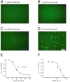
Doxazosin causes apoptosis of HL-1 cells, a cardiac cell line derived from a mouse atrial myocyte tumour lineage. (A–D) Fluorescence microphotographs corresponding to TUNEL assays. A total of 250 000 cells were employed per assay. Increased green nuclear fluorescence reflects endonucleolytic DNA degradation and apoptosis of cells treated with doxazosin. Scale bar, 100 μm. (E) Concentration–response relationship obtained from three independent XTT cell viability assays. (F) Time course of doxazosin-associated apoptosis. Half-maximal pro-apoptotic effects of 30 μM doxazosin were observed after 19.5 h (n = 5). Data are given as mean ± SEM.
Figure 2
Figure 2
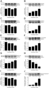
Analysis of proteins involved in pro-apoptotic signalling. (A, C) Total protein levels of EphA2 (A) and SHP-2 (C) were not significantly affected by doxazosin. In contrast, apoptosis was associated with increased phosphorylation (i.e. activation) of EphA2 (B), SHP-2 (D) and p38 MAPK (F). FAK exhibited reduced expression owing to cleavage (E), and growth arrest and DNA damage inducible gene 153 (GADD153) protein levels were elevated (G). (H) The anti-apoptotic protein kinase B (p-Akt) was suppressed. (I, J) Activation of caspase 3 en route to apoptosis. Doxazosin treatment induced cleavage of caspase 3 (I) associated with decreased expression of pre-processed caspase 3 levels (J) that did not reach statistical significance. Representative Western blots and mean ( ± SEM) optical densities normalized to doxazosin-free conditions are presented for HL-1 cells exposed to increasing concentrations of doxazosin (n = 3–5 independent assays; *P < 0.05; **P < 0.01). See text and Figure 5 for mechanistic details.
Figure 3
Figure 3
LCA prevents apoptosis. (A, B) Microscopic findings after treatment of HL-1 cells with vehicle (A) or 30 μM doxazosin (B) illustrate doxazosin-associated cell death. (C, D) LCA reduced (C) or prevented (D) apoptosis of cardiac myocytes (scale bar, 100 μm). (E) Quantification of mean cell survival normalized to controls using the XTT assay revealed dose-dependent apoptosis protection by LCA (n = 3 assays). (F, G) Activation of anti-apoptotic EphA2 signalling by LCA is illustrated by Western blot analyses of phosphorylated (F; n = 3–4 assays) and total EphA2 protein (G; n = 4 assays), respectively, revealing reduced phosphorylation and increased expression of EphA2 during doxazosin administration. LCA did not affect cells grown in the absence of the pro-apoptotic stimulus, doxazosin. Protein samples obtained from n = 4 assays was analysed per group. Data are provided as mean ± SEM [*P < 0.05; ***P < 0.001; ns, not significant vs. control medium (E) or doxazosin-free conditions (F, G) respectively].
Figure 4
Figure 4
Knock-down of EphA2 impairs cell viability. (A, B) Representative microphotographs illustrate reduced viability of HL-1 cells treated with siRNA to suppress EphA2 expression compared with controls exposed to scrambled siRNA (scale bar, 100 μm). (C) Quantification of viable cells normalized to controls. Cell survival was determined using an XTT-based assay (n = 6 assays; ***P < 0.001). Data are given as mean ± SEM. Scr, srambled siRNA.
Figure 5
Figure 5
EphA2 signalling in cardiac myocytes. Doxazosin triggers EphA2 phosphorylation, resulting in SHP-2 phosphorylation. FAK was inactivated. In addition, dephosphorylation of protein kinase B (Akt), phosphorylation of p38 MAPK, stimulation of growth arrest and DNA damage inducible gene 153 (GADD153) and activation (i.e. cleavage) of caspase 3 occurred. LCA inhibits apoptosis in the presence of doxazosin through prevention of EphA2 phosphorylation and via increased EphA2 protein expression. Note that, for clarity, intermediate signalling steps are not shown.

Similar articles

Cited by

References

    1. Aimé-Sempé C, Folliguet T, Rücker-Martin C, Krajewska M, Krajewska S, Heimburger M, et al. Myocardial cell death in fibrillating and dilated human right atria. J Am Coll Cardiol. 1999;34:1577–1586. - PubMed
    1. Alexander SP, Mathie A, Peters JA. Guide to Receptors and Channels (GRAC), 5th edition. Br J Pharmacol. 2011;164(Suppl. 1):S1–S324. - PMC - PubMed
    1. Bikou O, Thomas D, Trappe K, Lugenbiel P, Kelemen K, Koch M, et al. Connexin 43 gene therapy prevents persistent atrial fibrillation in a porcine model. Cardiovasc Res. 2011;92:218–225. - PubMed
    1. Brantley-Sieders DM, Zhuang G, Hicks D, Fang WB, Hwang Y, Cates JM, et al. The receptor tyrosine kinase EphA2 promotes mammary adenocarcinoma tumorigenesis and metastatic progression in mice by amplifying ErbB2 signaling. J Clin Invest. 2008;118:64–78. - PMC - PubMed
    1. Cardin S, Li D, Thorin-Trescases N, Leung TK, Thorin E, Nattel S. Evolution of the atrial fibrillation substrate in experimental congestive heart failure: angiotensin-dependent and -independent pathways. Cardiovasc Res. 2003;60:315–325. - PubMed

Publication types