Review
. 2013 May;169(2):304-17.
doi: 10.1111/j.1476-5381.2012.02118.x.

The case for induced pluripotent stem cell-derived cardiomyocytes in pharmacological screening

Affiliations
Review

The case for induced pluripotent stem cell-derived cardiomyocytes in pharmacological screening

Jaffar M Khan et al. Br J Pharmacol. 2013 May.

Abstract

The current drug screening models are deficient, particularly in detecting cardiac side effects. Human stem cell-derived cardiomyocytes could aid both early cardiotoxicity detection and novel drug discovery. Work over the last decade has generated human embryonic stem cells as potentially accurate sources of human cardiomyocytes, but ethical constraints and poor efficacy in establishing cell lines limit their use. Induced pluripotent stem cells do not require the use of human embryos and have the added advantage of producing patient-specific cardiomyocytes, allowing both generic and disease- and patient-specific pharmacological screening, as well as drug development through disease modelling. A critical question is whether sufficient standards have been achieved in the reliable and reproducible generation of 'adult-like' cardiomyocytes from human fibroblast tissue to progress from validation to safe use in practice and drug discovery. This review will highlight the need for a new experimental system, assess the validity of human induced pluripotent stem cell-derived cardiomyocytes and explore what the future may hold for their use in pharmacology.


Figures

Figure 1
Figure 1
Immunofluorescence image showing differentiated human induced pluripotent stem cell-derived cardiomyocytes. Cells are stained positive for cardiac-specific atrial natriuretic factor (cytoplasmic), mitotic marker Ki67 (nuclear) and myosin heavy chain a/β at 30 days after differentiation. Nuclei are stained with DAPI. Courtesy Gabor Foldes, NHLI, Imperial College.
Figure 2
Figure 2
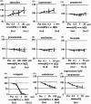
The effects of cardioactive drugs on the beating rates of contractile colonies derived from human iPS cells and human ES cells. Adrenaline, isoproterenol, verapamil, amiodarone and isoproterenol + propranolol had statistically significant effects between pre-drug loading; and the maximum concentration of the drug used in cardiomyocytes was derived from human iPS cells (P < 0.05). There were no statistically significant differences between the concentrations of drugs that elicited effects in human iPS cells and those that elicited effects in human ES cells (Yokoo et al. 2009, Biochem Biophys Res Commun 387: 482–488, with permission).
Figure 3
Figure 3
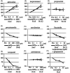
The effects of cardioactive drugs on the contractility of contractile colonies derived from human iPS cells and human ES cells. Adrenaline, isoproterenol, procainamide, flecainide, verapamil and isoproterenol + propranolol had statistically significant effects on human iPS cells between pre-drug loading and the maximum concentration of the drug used in cardiomyocytes derived from human iPS cells (P < 0.05). There were no statistically significant differences between the concentrations of drugs that elicited effects in human iPS cells and those that elicited effects in human ES cells (Yokoo et al. 2009, Biochem Biophys Res Commun 387: 482–488, with permission).
Figure 4
Figure 4
Action potential morphologies from iPSC-derived cardiomyocytes. From Ma J et al. Am J Physiol Heart Circ Physiol 2011;301: H2006–H2017 with permission. Interval, beat rate interval; MDP, maximum diastolic potential; Peak, peak voltage; Amp, amplitude; dV/dtmax, maximal rate of depolarization, APD, AP duration at different levels of repolarization (APD measured at 10% increments of Amp).
Figure 5
Figure 5
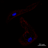
iCell-CMs (Cellular Dynamics International) were cultured for 2 weeks in iCell cardiomyocytes maintenance medium (Cellular Dynamics International), fixed with paraformaldehyde (4%) and immunolabelled with primary antibodies against mouse sarcomeric α-actinin (Sigma, A7811, 1:150). The secondary antibody was Alexa Fluor 594 goat anti mouse IgG (Invitrogen, 1:800). Nuclei were stained with DAPI. Scale bars: 20 µm. Courtesy Ljudmila Kolker, NISBC and Imperial College.

References

    1. Abdul Kadir SH, Ali NN, Mioulane M, Brito-Martins M, Abu-Hayyeh S, Foldes G, et al. Embryonic stem cell-derived cardiomyocytes as a model to study fetal arrhythmia related to maternal disease. J Cell Mol Med. 2009;13:3730–3741. - PMC - PubMed
    1. Avilion AA, Nicolis SK, Pevny LH, Perez L, Vivian N, Lovell-Badge R. Multipotent cell lineages in early mouse development depend on SOX2 function. Genes Dev. 2003;17:126–140. - PMC - PubMed
    1. Behringer RR. Human-animal chimeras in biomedical research. Cell Stem Cell. 2007;1:259–262. - PubMed
    1. Bers DM. Cardiac excitation-contraction coupling. Nature. 2002;415:198–205. - PubMed
    1. Binah O, Dolnikov K, Sadan O, Shilkrut M, Zeevi-Levin N, Amit M, et al. Functional and developmental properties of human embryonic stem cells-derived cardiomyocytes. J Electrocardiol. 2007;40(Suppl.):S192–S196. - PubMed