. 2012 Nov 1;21(21):4645-54.
doi: 10.1093/hmg/dds306. Epub 2012 Jul 30.

Mouse model of muscleblind-like 1 overexpression: skeletal muscle effects and therapeutic promise

Affiliations

Mouse model of muscleblind-like 1 overexpression: skeletal muscle effects and therapeutic promise

Christopher M Chamberlain et al. Hum Mol Genet. .

Abstract

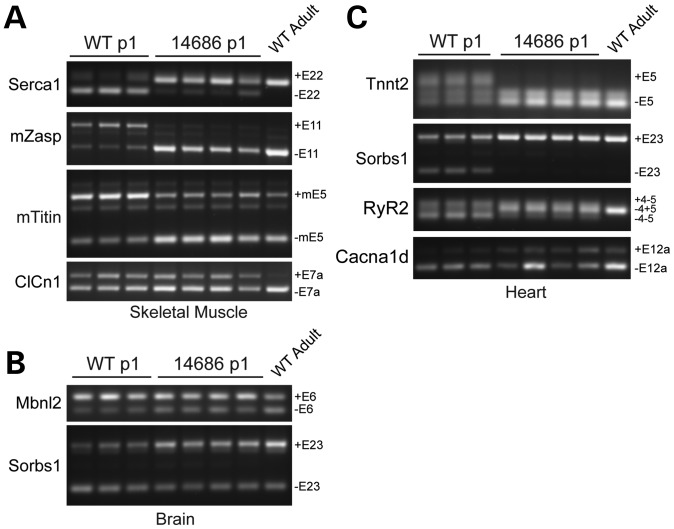
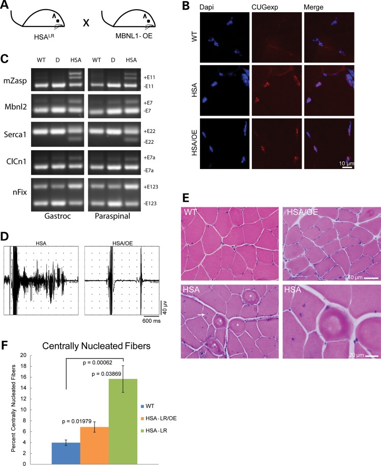
Myotonic dystrophy (DM) is a multisystemic disease caused by CTG or CCTG expansion mutations. There is strong evidence that DM1 CUG and DM2 CCUG expansion transcripts sequester muscleblind-like (MBNL) proteins and that loss of MBNL function causes alternative splicing abnormalities that contribute to disease. Because MBNL1 loss is thought to play an important role in disease and localized AAV delivery of MBNL1 partially rescues skeletal muscle pathology in DM mice, there is strong interest in MBNL1 overexpression as a therapeutic strategy. We developed the first transgenic MBNL1 overexpression mouse model (MBNL1-OE) to test the safety and efficacy of multisystemic MBNL1 overexpression. First, we demonstrate that MBNL1 overexpression is generally well-tolerated in skeletal muscle. Second, we show the surprising result that premature shifts in alternative splicing of MBNL1-regulated genes in multiple organ systems are compatible with life and do not cause embryonic lethality. Third, we show for the first time that early and long-term MBNL1 overexpression prevents CUG-induced myotonia, myopathy and alternative splicing abnormalities in DM1 mice. In summary, MBNL1 overexpression may be a valuable strategy for treating the skeletal muscle features of DM.


Figures

Figure 1.
Figure 1.
Transgene design and function. (A) Schematic diagram of the MBNL1-OE construct used to express the 40 kDa isoform, (−) exons 7 and 9, of human MBNL1 protein with a triple flag epitope on the N-terminus. Chicken β-actin promoter with early CMV enhancer (CAGG) drives transgene expression. (B) HEK293T cells transfected with 2 µg of Tnnt3 minigene plus 2 µg of either pcDNA3.1 empty vector, CUGBP1 (16) or MBNL1-OE vectors. Splicing of the Tnnt3 minigene was assayed using primers MSS1956 and MSS1938 (16).
Figure 2.
Figure 2.
Recombinant MBNL1 expression and localization in MBNL1-OE mice. (A) Western blots of tissues from FVB, 14685 and 14686 mice at 6 months of age. Recombinant MBNL1 and endogenous Mbnl1 are detected using A2764 (αMbnl1) primary and HRP-conjugated α-rabbit secondary antibodies. αFlag-HRP (M2) and α-mouse-HRP detect recombinant MBNL1 only. GAPDH was used as a loading control. (B) Western blots of whole embryo protein lysates show expression of recombinant and endogenous MBNL1/Mbnl1 for 14685 at embryonic day 11 (E11) and 14686 at E13. (C) Relative levels of recombinant (MBNL1) and endogenous (Mbnl1) protein in 14685 (n = 7) versus wild-type littermates (n = 3) and 14686 (n = 3) versus wild-type littermates (n = 3) were quantified and normalized to the alpha-tubulin loading control. Endogenous Mbnl1 levels in wild-type controls were set to 1 to calculate a fold-change in total MBNL1/Mbnl1 levels. *P < 0.05; **P < 0.005; ***P < 0.001. (D) Immunofluorescence of recombinant MBNL1 in 14685 gastrocnemius muscle using α-Flag (F7425 Sigma) and α-rabbit Alexa Fluor 488 (Jackson Immunoresearch) antibodies. No recombinant protein is detected in wild-type controls. DAPI stain was used to detect nuclei.
Figure 3.
Figure 3.
Lifespan and mass of MBNL1-OE mice. (A) Kaplan–Meier survival plot for 14685 (n = 35 WT; n = 28 OE) and (B) 14686 (n = 26 WT; n = 23 OE) mice up to 78 weeks of age. No statistically significant difference in survival at any time point was observed in line 14685. Mice from line 14686 have a significant increase in percent mortality beginning at week 76 (P = 0.023). (C) Mass of 14685 and (D) 14686 mice (n >3 for all time points). No significant difference in mass was observed for line 14685 at any time point. In contrast, 14686 mice were smaller at 3–8 weeks (P < 0.005) and for week 9 (P = 0.011). Asterisks (*) indicate statistically significant differences.
Figure 4.
Figure 4.
Muscle histology and function in MBNL1-OE mice. (A) Hematoxylin and eosin stains of skeletal muscle from 14685, 14686 and wild-type littermate controls. (B) Percentage of fibers with at least one central nuclei in skeletal muscle from wild-type (n = 6), 14685 (n = 4) and 14686 (n = 3) animals at 11–14 months. (C) Average fiber cross-sectional area in 11–14-month wild-type (n = 6), 14685 (n = 4) and 14686 (n = 3) mice. (D) Total forelimb grip strength in 4–7-month wild-type (n = 21), 14685 (n = 15) and 14686 (n = 11) mice. 14686 have a significant loss in forelimb grip strength compared with wild-type controls (P < 0.001). (E) Grip strength normalized to skeletal muscle mass in 4–7-month wild-type (n = 5) and 14686 (n = 5) mice. (F) Rotarod analysis at 6 months in 14685 (n = 10) versus wild-type littermates (n = 7) and 14686 (n = 8) versus wild-type littermates (n = 8).
Figure 5.
Figure 5.
Neonatal splicing in MBNL1-OE mice. RT-PCR analysis of alternative splicing in 14686 and wild-type littermates at post-natal day 1. mRNA targets and specific alternatively spliced exons are listed on each gel. Mbnl1 targets were analyzed in skeletal muscle (A), brain (B) and heart (C).
Figure 6.
Figure 6.
MBNL1 overexpression corrects a DM skeletal muscle phenotype. (A) Schematic diagram of MBNL1-OE and HSALR cross. (B) RNA foci in skeletal muscle from HSALR and HSALR;MBNL1-OE mice detected with a Cy3 conjugated CAG8 oligonucleotide probe. No foci were detected in wild-type FVB mice. (C) RT-PCR splicing assays of FVB wild-type (WT), singly transgenic HSALR (HSA) and doubly transgenic HSALR;MBNL1-OE mice (D). (D) EMG trace from an HSALR gastrocnemius muscle showing a single needle insertion resulting in a ∼2s myotonic discharge and trace from an HSALR;MBNL1-OE doubly transgenic mouse gastrocnemius showing two normal needle insertion responses without electrical myotonia. (E) Hematoxylin and eosin staining of quadriceps muscle from 12-month FVB wild-type, singly transgenic HSALR and doubly transgenic HSALR;MBNL1-OE mice. Ringed fibers (asterisk) and multiple central nuclei (arrow) shown in the HSALR mouse. (F) Quantitation of centrally nucleated fibers in FVB wild-type, HSALR and HSALR;MBNL1-OE mice. Averages are shown ±SEM.

References

    1. Harper P.S. Myotonic Dystrophy. 3rd edn. London: W.B. Saunders; 2001.
    1. Liquori C.L., Ricker K., Moseley M.L., Jacobsen J.F., Kress W., Naylor S.L., Day J.W., Ranum L.P. Myotonic dystrophy type 2 caused by a CCTG expansion in intron 1 of ZNF9. Science. 2001;293:864–867. - PubMed
    1. Brook J.D., McCurrach M.E., Harley H.G., Buckler A.J., Church D., Aburatani H., Hunter K., Stanton V.P., Thirion J.P., Hudson T. Molecular basis of myotonic dystrophy: expansion of a trinucleotide (CTG) repeat at the 3′ end of a transcript encoding a protein kinase family member. Cell. 1992;69:385. - PubMed
    1. Mahadevan M., Tsilfidis C., Sabourin L., Shutler G., Amemiya C., Jansen G., Neville C., Narang M., Barceló J., O'Hoy K. Myotonic dystrophy mutation: an unstable CTG repeat in the 3′ untranslated region of the gene. Science. 1992;255:1253–1255. - PubMed
    1. Fu Y.H., Pizzuti A., Fenwick R.G., King J., Rajnarayan S., Dunne P.W., Dubel J., Nasser G.A., Ashizawa T., de Jong P. An unstable triplet repeat in a gene related to myotonic muscular dystrophy. Science. 1992;255:1256–1258. - PubMed

Publication types