

Regional changes in thalamic shape and volume with increasing age
- PMID: 22846656
- PMCID: PMC3507623
- DOI: 10.1016/j.neuroimage.2012.07.043
Regional changes in thalamic shape and volume with increasing age
Abstract
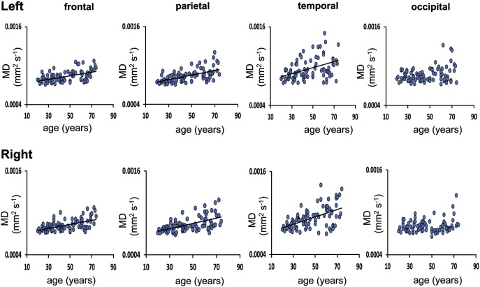
The thalamus undergoes significant volume loss and microstructural change with increasing age. Alterations in thalamo-cortical connectivity may contribute to the decline in cognitive ability associated with aging. The aim of this study was to assess changes in thalamic shape and in the volume and diffusivity of thalamic regions parcellated by their connectivity to specific cortical regions in order to test the hypothesis age related thalamic change primarily affects thalamic nuclei connecting to the frontal cortex. Using structural magnetic resonance imaging (MRI) and diffusion tensor imaging (DTI), we assessed thalamic volume and diffusivity in 86 healthy volunteers, median (range) age 44 (20-74) years. Regional thalamic micro and macro structural changes were assessed by segmenting the thalamus based on connectivity to the frontal, parietal, temporal and occipital cortices and determining the volumes and mean diffusivity of the thalamic projections. Linear regression analysis was performed to test the relationship between increasing age and (i) normalised thalamic volume, (ii) whole thalamus diffusion measures, (iii) mean diffusivity (MD) of the thalamo-cortical projections, and (iv) volumes of the thalamo-cortical projections. We also assessed thalamic shape change using vertex analysis. We observed a significant reduction in the volume and a significant increase in MD of the whole thalamus with increasing age. The volume of the thalamo-frontal projections decreased significantly with increasing age, however there was no significant relationship between the volumes of the thalamo-cortical projections to the parietal, temporal, and occipital cortex and age. Thalamic shape analysis showed that the greatest shape change was in the anterior thalamus, incorporating regions containing the anterior nucleus, the ventroanterior nucleus and the dorsomedial nucleus. To explore these results further we studied two additional groups of subjects (a younger and an older aged group, n=20), which showed that the volume of the thalamo-frontal projections was correlated to executive functions scores, as assessed by the Stroop test. These data suggest that atrophy of the frontal thalamo-cortical unit may explain, at least in part, disorders of attention, working memory and executive function associated with increasing age.
Copyright © 2012 Elsevier Inc. All rights reserved.
Figures


Similar articles
-
Multiple thalamo-cortical disconnections in anterior thalamic infarction: implications for thalamic mechanisms of memory and language.Neuropsychologia. 2014 Jan;53:264-73. doi: 10.1016/j.neuropsychologia.2013.11.025. Epub 2013 Dec 7. Neuropsychologia. 2014. PMID: 24321272
-
Thalamo-cortical network pathology in idiopathic generalized epilepsy: insights from MRI-based morphometric correlation analysis.Neuroimage. 2009 Jun;46(2):373-81. doi: 10.1016/j.neuroimage.2009.01.055. Neuroimage. 2009. PMID: 19385011
-
Medio-dorsal thalamus and confabulations: Evidence from a clinical case and combined MRI/DTI study.Neuroimage Clin. 2016 Oct 12;12:776-784. doi: 10.1016/j.nicl.2016.10.011. eCollection 2016. Neuroimage Clin. 2016. PMID: 27812504 Free PMC article.
-
Thalamic structures and associated cognitive functions: Relations with age and aging.Neurosci Biobehav Rev. 2015 Jul;54:29-37. doi: 10.1016/j.neubiorev.2015.03.008. Epub 2015 Apr 9. Neurosci Biobehav Rev. 2015. PMID: 25862940 Free PMC article. Review.
-
Subcortical Alterations in Newly Diagnosed Epilepsy and Associated Changes in Brain Connectivity and Cognition.Hum Brain Mapp. 2024 Nov;45(16):e70069. doi: 10.1002/hbm.70069. Hum Brain Mapp. 2024. PMID: 39508641 Free PMC article. Review.
Cited by
-
The Neurochemical and Microstructural Changes in the Brain of Systemic Lupus Erythematosus Patients: A Multimodal MRI Study.Sci Rep. 2016 Jan 13;6:19026. doi: 10.1038/srep19026. Sci Rep. 2016. PMID: 26758023 Free PMC article.
-
Structure of subcortico-cortical tracts in middle-aged and older adults with autism spectrum disorder.Cereb Cortex. 2024 Dec 3;34(12):bhae457. doi: 10.1093/cercor/bhae457. Cereb Cortex. 2024. PMID: 39707985
-
Brain Volume Metric Analysis Is Correlated with Aging Changes and Sex Differences in Thai Older Adults.Dement Geriatr Cogn Dis Extra. 2025 Jan 27;15(1):47-57. doi: 10.1159/000543774. eCollection 2025 Jan-Dec. Dement Geriatr Cogn Dis Extra. 2025. PMID: 40093354 Free PMC article.
-
Toward a Common Terminology for the Thalamus.Front Neuroanat. 2019 Jan 11;12:114. doi: 10.3389/fnana.2018.00114. eCollection 2018. Front Neuroanat. 2019. PMID: 30687023 Free PMC article. Review.
-
Relationship among interthalamic adhesion size, thalamic anatomy and neuropsychological functions in healthy volunteers.Brain Struct Funct. 2017 Jul;222(5):2183-2192. doi: 10.1007/s00429-016-1334-6. Epub 2016 Nov 19. Brain Struct Funct. 2017. PMID: 27866270 Free PMC article.
References
-
- Abe O., Yamasue H., Aoki S., Suga M., Yamada H., Kasai K., Masutani Y., Kato N., Ohtomo K. Aging in the CNS: comparison of gray/white matter volume and diffusion tensor data. Neurobiol. Aging. 2008;29:102–116. - PubMed
-
- Behrens T.E., Johansen-Berg H., Woolrich M.W., Smith S.M., Wheeler-Kingshott C.A., Boulby P.A., Barker G.J., Sillery E.L., Sheehan K., Ciccarelli O. Non-invasive mapping of connections between human thalamus and cortex using diffusion imaging. Nat. Neurosci. 2003;6:750–757. - PubMed
-
- Channon S. Frontal lobe dysfunction and everyday problem-solving: social and non-social contributions. Acta Psychol. (Amst) 2004;115:235–254. - PubMed
Publication types
MeSH terms
Grants and funding
LinkOut - more resources
Full Text Sources
Medical
