Review
. 2013 Feb;66(2):469-78.
doi: 10.1016/j.ympev.2012.07.013. Epub 2012 Jul 27.

Gene duplication, genome duplication, and the functional diversification of vertebrate globins

Affiliations
Review

Gene duplication, genome duplication, and the functional diversification of vertebrate globins

Jay F Storz et al. Mol Phylogenet Evol. 2013 Feb.

Abstract

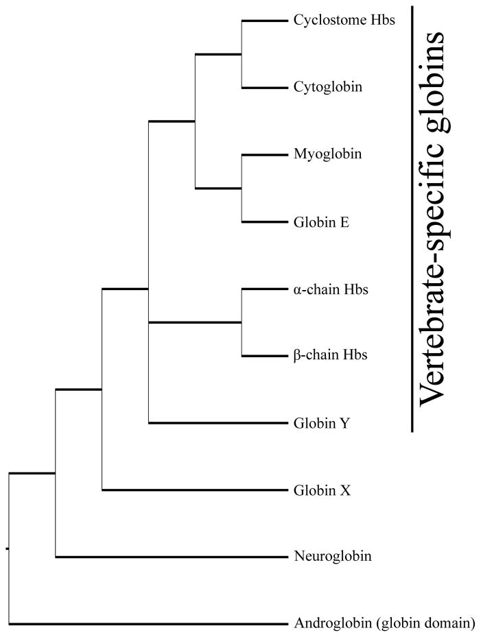
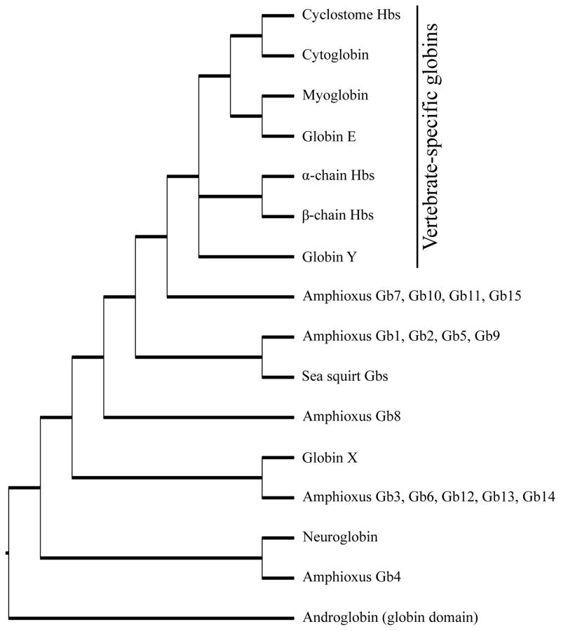
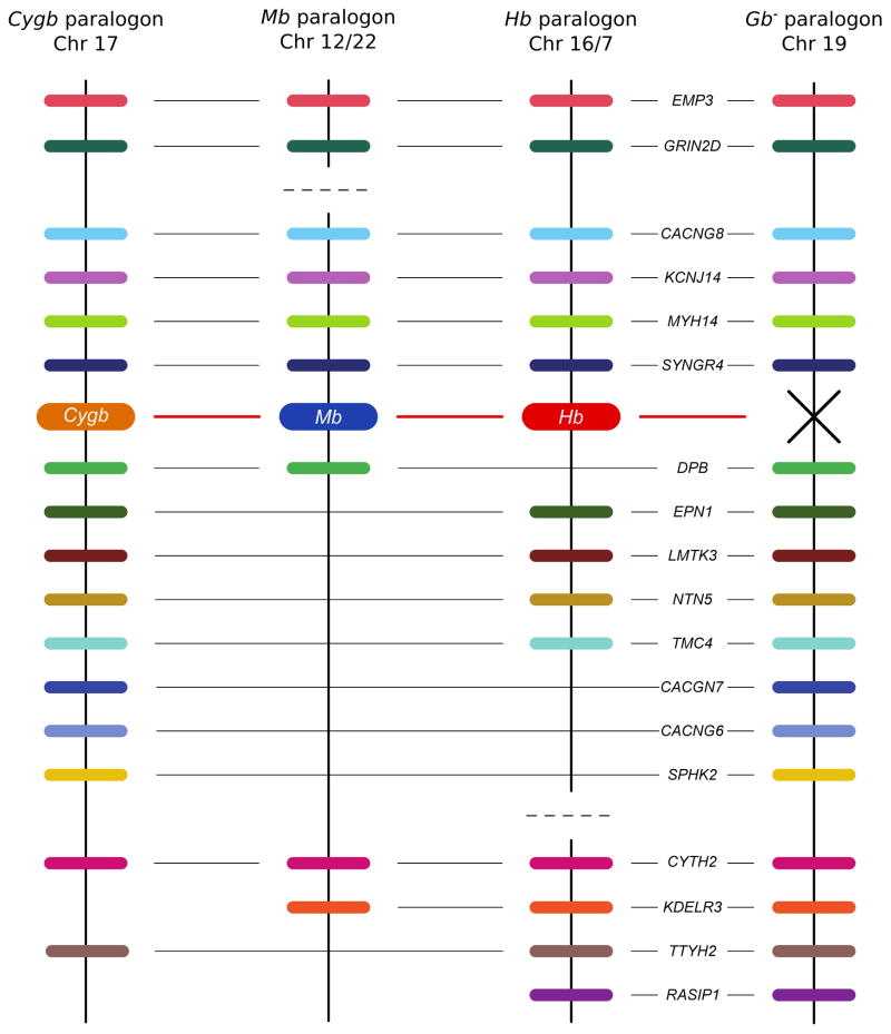
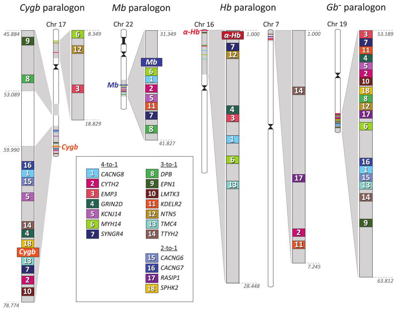
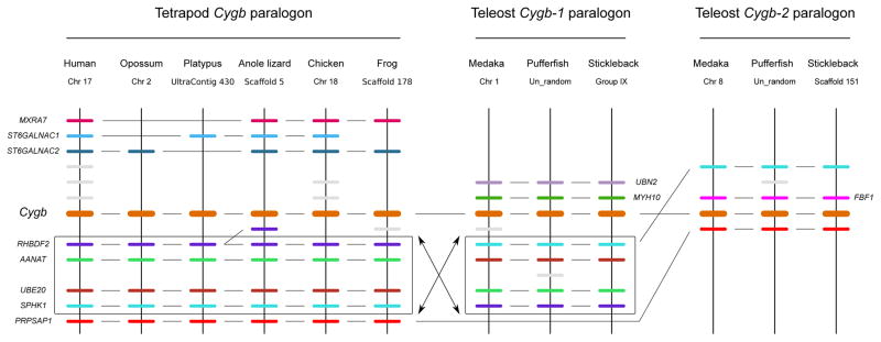
The functional diversification of the vertebrate globin gene superfamily provides an especially vivid illustration of the role of gene duplication and whole-genome duplication in promoting evolutionary innovation. For example, key globin proteins that evolved specialized functions in various aspects of oxidative metabolism and oxygen signaling pathways (hemoglobin [Hb], myoglobin [Mb], and cytoglobin [Cygb]) trace their origins to two whole-genome duplication events in the stem lineage of vertebrates. The retention of the proto-Hb and Mb genes in the ancestor of jawed vertebrates permitted a physiological division of labor between the oxygen-carrier function of Hb and the oxygen-storage function of Mb. In the Hb gene lineage, a subsequent tandem gene duplication gave rise to the proto α- and β-globin genes, which permitted the formation of multimeric Hbs composed of unlike subunits (α(2)β(2)). The evolution of this heteromeric quaternary structure was central to the emergence of Hb as a specialized oxygen-transport protein because it provided a mechanism for cooperative oxygen-binding and allosteric regulatory control. Subsequent rounds of duplication and divergence have produced diverse repertoires of α- and β-like globin genes that are ontogenetically regulated such that functionally distinct Hb isoforms are expressed during different stages of prenatal development and postnatal life. In the ancestor of jawless fishes, the proto Mb and Hb genes appear to have been secondarily lost, and the Cygb homolog evolved a specialized respiratory function in blood-oxygen transport. Phylogenetic and comparative genomic analyses of the vertebrate globin gene superfamily have revealed numerous instances in which paralogous globins have convergently evolved similar expression patterns and/or similar functional specializations in different organismal lineages.


Figures

Figure 1
Figure 1
Cladogram describing phylogenetic relationships among vertebrate globins.
Figure 2
Figure 2
Oxygen equilibrium curves for human Mb and Hb in whole blood. The dashed curve is a hyperbolic oxygen equilibrium curve with the same P50 (the partial pressure of oxygen at which Hb is half saturated) as human Hb (26 torr).
Figure 3
Figure 3
Cladogram describing phylogenetic relationships among members of the globin gene superfamily in chordates.
Figure 4
Figure 4
Diagram depicting the hypothetical effects of two consecutive genome duplications (1R and 2R), as reflected in the physical linkage arrangement of paralogous genes – specifically, the four-fold pattern of intragenomic synteny. (A) Hypothetical chromosome in the vertebrate common ancestor; (B) The first genome duplication produces a complete set of paralogs in identical order; (C) Many paralogous gene copies are subsequently deleted from the genome; (D) The second genome duplication produces yet another set of paralogs in identical order, with multigene families that retained two copies now present in four. (E) With the passage of time, additional gene losses ensue, thereby obscuring the four-fold pattern of synteny. Modified from Dehal and Boore (2005).
Figure 5
Figure 5
Graphical depiction of gene duplicates that are shared between the Gb paralogon and the remaining three globin-defined paralogons (Cygb, Mb, and Hb) in the human genome. There are seven 4:1 gene families that unite the Gb paralogon with the Cygb, Mb, and Hb paralogons, there are seven 3:1 gene families that unite the Gb paralogon with two of the three globin-defined paralogons, and there are four 2:1 gene families that unite the Gb paralogon with a single globin-defined paralogon. On each chromosome, annotated genes are depicted as colored bars. The ‘missing’ globin gene on the Gb paralogon is denoted by an ‘X’. The shared paralogs are depicted in colinear arrays for display purposes only, as there is substantial variation in gene order among the four paralogons. For clarity of presentation, genes that are not shared between the Gb paralogon and any of the three globin-defined paralogons are not shown. In the human genome, the Gb paralogon on chromosome 19 shares multiple gene duplicates with fragments of the Hb paralogon on chromosomes 16 and 7, and fragments of the Mb paralogon on Chromosomes 12 and 22. Members of the EPN1, LMTK3, and KCNJ14 gene families that map to the Hb paralogon have been secondarily translocated from chromosome 16. From Hoffmann et al. (2012a).
Figure 6
Figure 6
Four-fold pattern of conserved macrosynteny between the four globin-defined paralogons in the human genome (including the Gb paralogon) and ‘linkage group 15’ of the reconstructed proto-karyotype of the chordate common ancestor (Putnam et al. 2008; shaded regions). This pattern of conserved macrosynteny demonstrates that the Cygb, Mb, Hb, and Gb paralogons trace their duplicative origins to the same proto-chromosome of the chordate common ancestor, and provides conclusive evidence that each of the four paralogons are products of a genome quadruplication in the stem lineage of vertebrates. Shared gene duplicates that map to secondarily translocated segments of the Mb paralogon (on chromosome 7) and the Hb paralogon (on chromosomes 12 and 17) are not pictured. From Hoffmann et al. (2012a).
Figure 7
Figure 7
Patterns of conserved synteny in the chromosomal region that harbors the Cygb gene in gnathostome vertebrates. Horizontal lines denote orthologous relationships. From Hoffmann et al. (2011).

References

    1. Abi-Rached L, Gilles A, Shiina T, Pontarotti P, Inoko H. Evidence of en bloc duplication in vertebrate genomes. Nat Genet. 2002;31:100–105. - PubMed
    1. Alev C, Shinmyozu K, McIntyre BAS, Sheng G. Genomic organization of zebra finch α and β globin genes and their expression in primitive and definitive blood in comparison with globins in chicken. Dev Genes Evol. 2009;219:353–360. - PubMed
    1. Awenius C, Hankeln T, Burmester T. Neuroglobins from the zebrafish Danio rerio and the pufferfish Tetraodon nigroviridis. Biochem Biophys Res Commun. 2001;287:418–421. - PubMed
    1. Blank M, Kiger L, Thielebein A, Gerlach F, Hankeln T, Marden MC, Burmester T. Oxygen supply from the bird’s eye perspective: Globin E is a respiratory protein in the chicken retina. J Biol Chem. 2011a;286:26507–26515. - PMC - PubMed
    1. Blank M, Wollberg J, Gerlach F, Reimann K, Roesner A, Hankeln T, Fago A, Weber RE, Burmester T. A membrane-bound vertebrate globin. PLoS ONE. 2011b;6:e25292. - PMC - PubMed

Publication types

LinkOut - more resources