. 2012 Oct 26;423(3):397-412.
doi: 10.1016/j.jmb.2012.07.017. Epub 2012 Jul 27.

Construction of a stability landscape of the CH3 domain of human IgG1 by combining directed evolution with high throughput sequencing

Affiliations

Construction of a stability landscape of the CH3 domain of human IgG1 by combining directed evolution with high throughput sequencing

Michael W Traxlmayr et al. J Mol Biol. .

Abstract

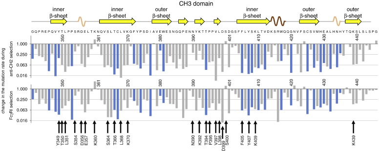
One of the most important but still poorly understood issues in protein chemistry is the relationship between sequence and stability of proteins. Here, we present a method for analyzing the influence of each individual residue on the foldability and stability of an entire protein. A randomly mutated library of the crystallizable fragment of human immunoglobulin G class 1 (IgG1-Fc) was expressed on the surface of yeast, followed by heat incubation at 79°C and selection of stable variants that still bound to structurally specific ligands. High throughput sequencing allowed comparison of the mutation rate between the starting and selected library pools, enabling the generation of a stability landscape for the entire CH3 domain of human IgG1 at single residue resolution. Its quality was analyzed with respect to (i) the structure of IgG1-Fc, (ii) evolutionarily conserved positions and (iii) in silico calculations of the energy of unfolding of all variants in comparison with the wild-type protein. In addition, this new experimental approach allowed the assignment of functional epitopes of structurally specific ligands used for selection [Fc γ-receptor I (CD64) and anti-human CH2 domain antibody] to distinct binding regions in the CH2 domain.


Figures

None
Graphical abstract
Fig. 1
Fig. 1
(a) The crystal structure of human IgG1-Fc (PDB ID 1OQO) is depicted using PyMOL. IgG1-Fc is composed of two polypeptide chains (dark and light gray, respectively), each of which comprises a CH2 domain (top) and a CH3 domain (bottom). The N-linked glycosylation is located between the two CH2 domains (shown in sticks). The hinge region with the two disulfide bonds connecting the two chains is missing in this structure. (b) Characterization of the original library and Fc-wt. An IgG1-Fc library was constructed by error-prone PCR, displayed on yeast and probed for binding to an antibody directed against an N-terminal expression tag (anti-Xpress) and to either anti-CH2 (upper row) or FcγRI (lower row). (c) Comparison of original and selected libraries. The yeast‐displayed Fc library was incubated at 79 °C for 10 min, followed by selection for retained binding to either FcγRI or anti-CH2. For comparison, the original library, the selected libraries and Fc-wt were displayed on yeast again, followed by heat incubation at 79 °C for 10 min. The samples were cooled and subsequently probed for binding to anti-Xpress and to either anti-CH2 (upper row) or FcγRI (lower row). In (b) and (c), only cells displaying the N-terminal tag (Xpress positive) are shown. The percentages of cells that are located in the gates (negative for binding to anti-CH2 or FcγRI) are indicated in the dot plots. For comparison, the gates were set identically in each row, respectively.
Fig. 2
Fig. 2
Stability/epitope landscape of the CH2 domain. The original library and the two selected libraries were analyzed by high throughput sequencing. The mutation rate at each amino acid position was calculated and compared between the selected and original libraries, yielding the change in the mutation rate during the selection process at each amino acid position. In this figure, mutation rate changes at all positions in the CH2 domain (also including the C-terminal part of the hinge region) during the anti-CH2 selection (upper diagram) and the FcγRI selection (lower diagram) are depicted. On the top, the secondary structural elements of the CH2 domain are shown (PDB ID 1OQO) (yellow, β-strand; dark brown, α-helix). Underneath the secondary structure, the wild-type sequence is shown, together with numbers indicating the amino acid positions (Eu numbering system17). Bars of evolutionarily conserved residues that are identical in the IgG1-CH2 domains of all 11 analyzed species are depicted in blue. Positions where the FcγRI/anti-CH2 ratio (change in the mutation rate during FcγRI selection divided by the one during anti-CH2 selection) is higher than 8 or lower than 1/8 are marked with green arrows (more intolerant to mutation during FcγRI selection) or orange arrows (more intolerant to mutation during anti-CH2 selection).
Fig. 3
Fig. 3
Stability landscape of the CH3 domain. Analog to Fig. 2, the stability landscape of the CH3 domain is depicted. The following secondary structural elements are shown in this figure (according to PDB ID 1OQO): yellow, β-strand; dark brown, α-helix; and light brown, 3/10-helix. In addition, β-strands building the inner and outer β-sheets are labeled. Bars of evolutionarily conserved residues that are identical in the IgG1-CH3 domains of all 12 analyzed species are depicted in blue. Amino acids that are located within 4 Å from the other CH3 domain are marked with black arrows.
Fig. 4
Fig. 4
(a) Amino acids of the CH3 domain are divided into two groups: residues that are evolutionarily conserved (identical in the CH3 domains of IgG1 of all 12 analyzed species) and those that are not. Subsequently, in both groups, the geometric mean of the mutation rate changes during the FcγRI and anti-CH2 selections was calculated and statistically compared by using the Student's t-Test (Microsoft Excel). (b) Amino acids of the CH3 domain are divided into two groups based on whether or not they are located at the CH3–CH3 interface of the homodimeric protein. Amino acids located within 4 Å from the other CH3 domain are considered as being part of the CH3–CH3 interface, whereas all other amino acids with a distance of more than 4 Å are not included in this group. Again, the average values of the changes in the mutation rates are shown and compared.
Fig. 5
Fig. 5
Comparison of the mutation rate change during selection and the predicted ΔΔG at positions located in the CH3 domain. ΔΔG values were calculated for all mutations. For each position, the median ΔΔG was calculated from all 19 possible changes and plotted against the mutation rate change during selection (geometric mean of the two experiments). Residues that are discussed in detail in the text are depicted in red.
Fig. 6
Fig. 6
Comparison of the mutation rate change during anti-CH2 and FcγRI selections. The changes in the mutation rates during the anti-CH2 and FcγRI selections are shown for each amino acid position in the CH2 (a) or CH3 (b) domain. Evolutionarily conserved residues that are identical in all analyzed species (11 species for the CH2 domain and 12 species for the CH3 domain) are depicted in blue.
Fig. 7
Fig. 7
Location of residues that are intolerant to mutation. The three‐dimensional structure [with secondary structural elements (left) or a space‐filling model (right)] of homodimeric human IgG1-Fc (PDB ID 1OQO) is shown using PyMOL. The glycosylation is shown in gray. Amino acids are colored according to their mutation rate change during anti-CH2 selection (top) or FcγRI selection (bottom). Residues that are involved in ligand binding (FcγRI/anti-CH2 ratio higher than 8 or lower than 1/8) are labeled with orange arrows.

Similar articles

Cited by

References

    1. Magliery T.J., Lavinder J.J., Sullivan B.J. Protein stability by number: high-throughput and statistical approaches to one of protein science's most difficult problems. Curr. Opin. Chem. Biol. 2011;15:443–451. - PMC - PubMed
    1. Hanes J., Pluckthun A. In vitro selection and evolution of functional proteins by using ribosome display. Proc. Natl Acad. Sci. USA. 1997;94:4937–4942. - PMC - PubMed
    1. He M., Taussig M.J. Antibody–ribosome–mRNA (ARM) complexes as efficient selection particles for in vitro display and evolution of antibody combining sites. Nucleic Acids Res. 1997;25:5132–5134. - PMC - PubMed
    1. McCafferty J., Griffiths A.D., Winter G., Chiswell D.J. Phage antibodies: filamentous phage displaying antibody variable domains. Nature. 1990;348:552–554. - PubMed
    1. Smith G.P. Filamentous fusion phage: novel expression vectors that display cloned antigens on the virion surface. Science. 1985;228:1315–1317. - PubMed

Publication types

Substances

LinkOut - more resources