Comparative Study
. 2012 Aug;73(2):391-400.
doi: 10.1097/TA.0b013e31825c7a82.

Early stabilizing alveolar ventilation prevents acute respiratory distress syndrome: a novel timing-based ventilatory intervention to avert lung injury

Affiliations
Comparative Study

Early stabilizing alveolar ventilation prevents acute respiratory distress syndrome: a novel timing-based ventilatory intervention to avert lung injury

Shreyas Roy et al. J Trauma Acute Care Surg. 2012 Aug.

Abstract

Background: Established acute respiratory distress syndrome (ARDS) is often refractory to treatment. Clinical trials have demonstrated modest treatment effects, and mortality remains high. Ventilator strategies must be developed to prevent ARDS.

Hypothesis: Early ventilatory intervention will block progression to ARDS if the ventilator mode (1) maintains alveolar stability and (2) reduces pulmonary edema formation.

Methods: Yorkshire pigs (38-45 kg) were anesthetized and subjected to a "two-hit" ischemia-reperfusion and peritoneal sepsis. After injury, animals were randomized into two groups: early preventative ventilation (airway pressure release ventilation [APRV]) versus nonpreventative ventilation (NPV) and followed for 48 hours. All animals received anesthesia, antibiotics, and fluid or vasopressor therapy as per the Surviving Sepsis Campaign. Titrated for optimal alveolar stability were the following ventilation parameters: (1) NPV group--tidal volume, 10 mL/kg + positive end-expiratory pressure - 5 cm/H2O volume-cycled mode; (2) APRV group--tidal volume, 10 to 15 mL/kg; high pressure, low pressure, time duration of inspiration (Thigh), and time duration of release phase (Tlow). Physiological data and plasma were collected throughout the 48-hour study period, followed by BAL and necropsy.

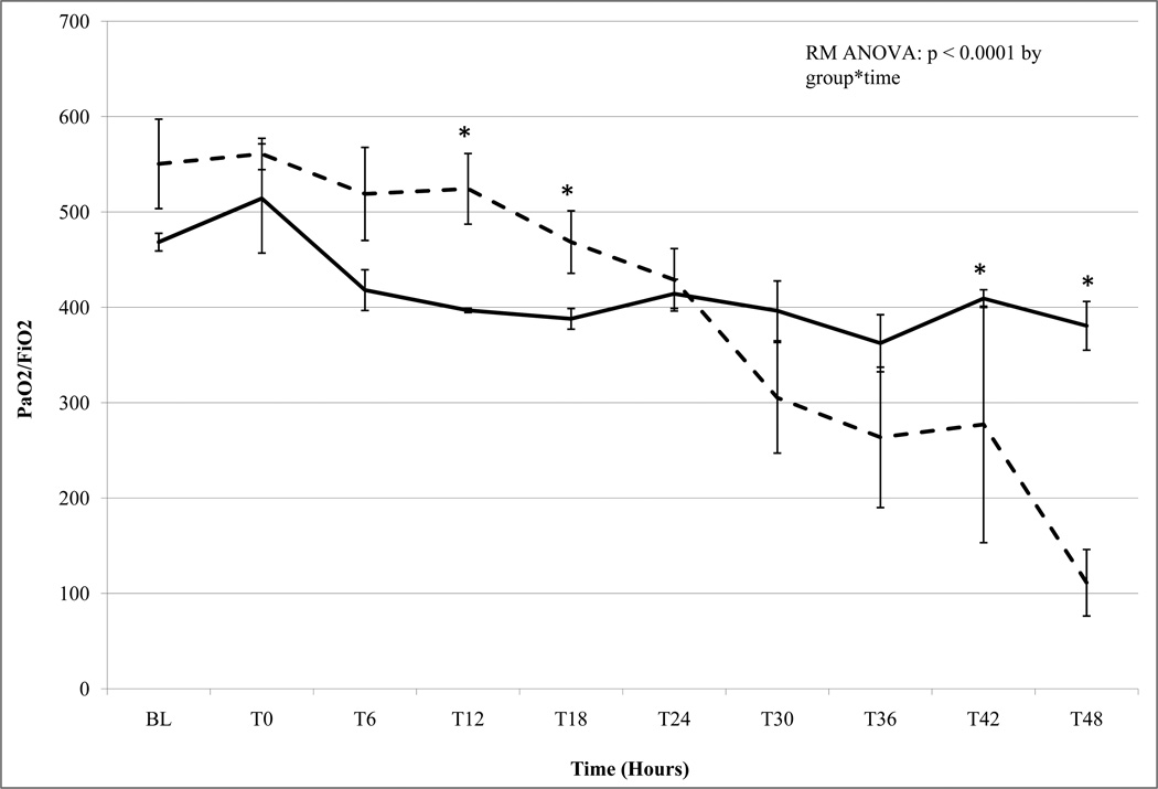
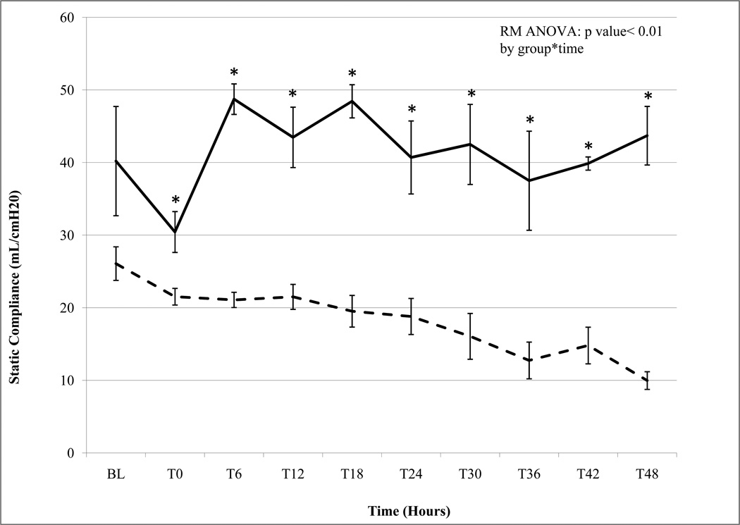
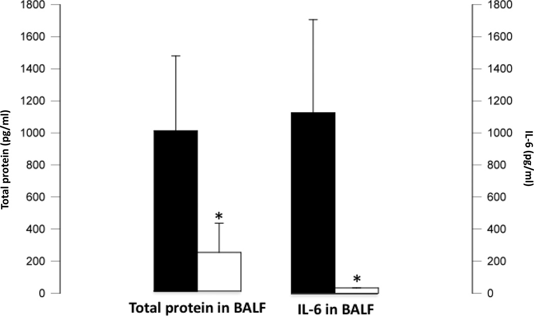
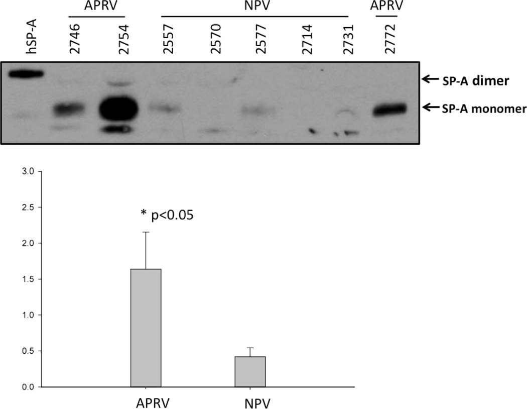
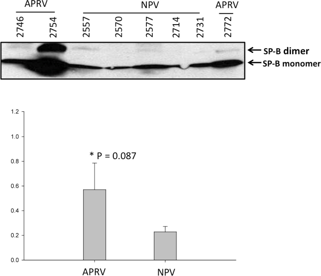
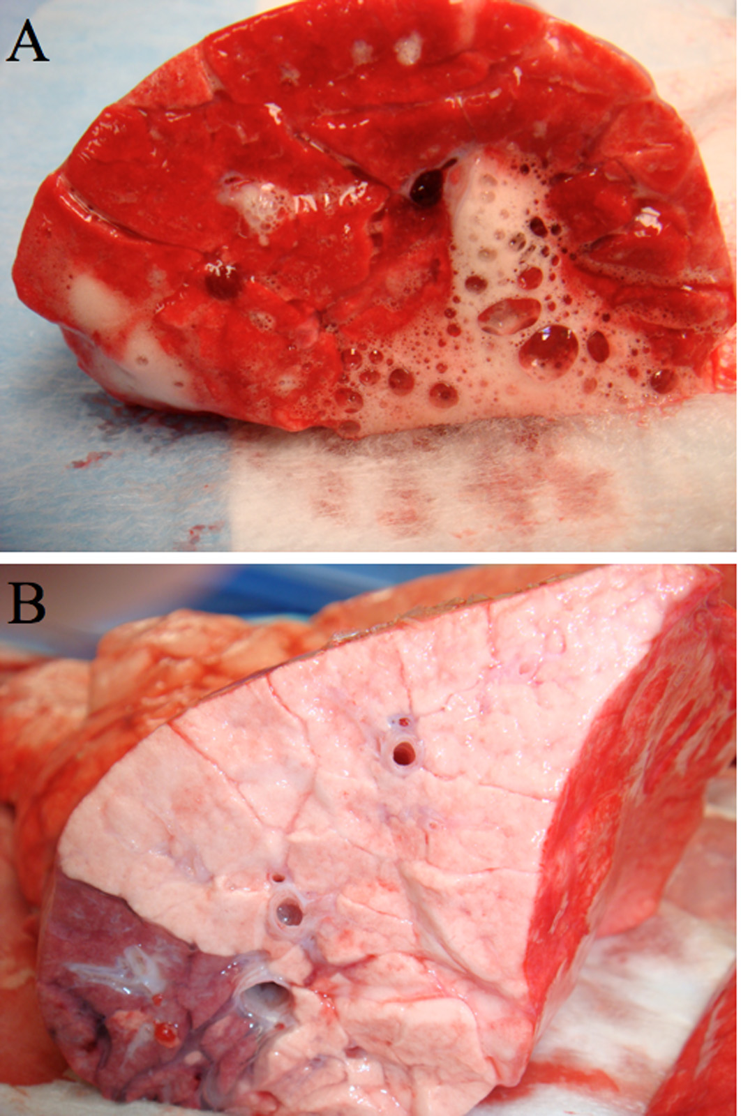
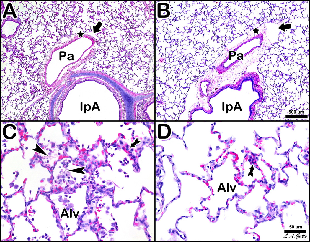
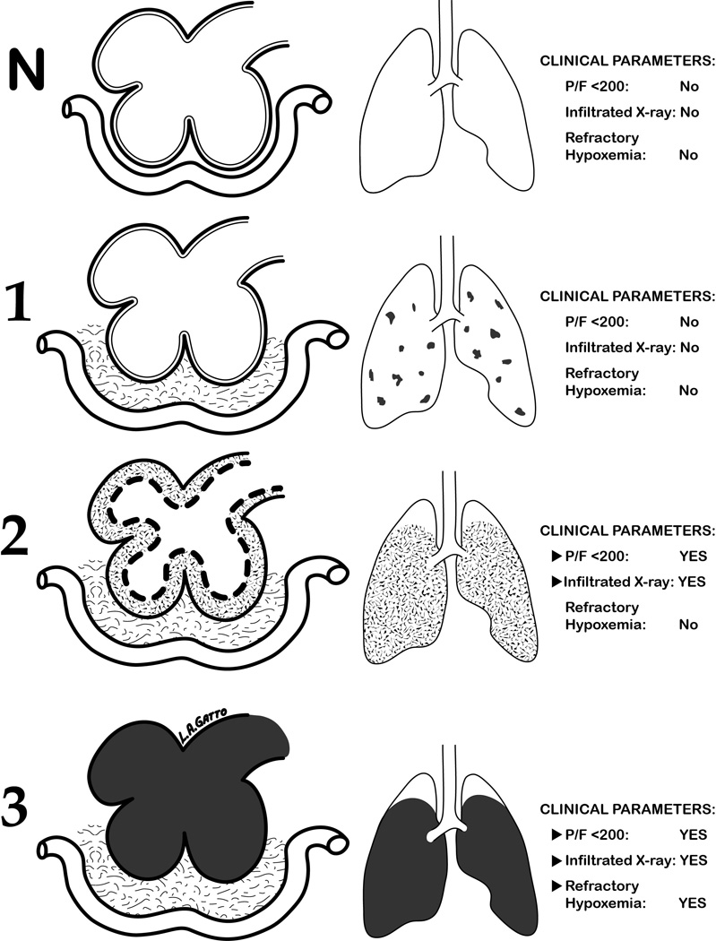
Results: APRV prevented the development of ARDS (p < 0.001 vs. NPV) by PaO₂/FIO₂ ratio. Quantitative histological scoring showed that APRV prevented lung tissue injury (p < 0.001 vs. NPV). Bronchoalveolar lavage fluid showed that APRV lowered total protein and interleukin 6 while preserving surfactant proteins A and B (p < 0.05 vs. NPV). APRV significantly lowered lung water (p < 0.001 vs. NPV). Plasma interleukin 6 concentrations were similar between groups.

Conclusion: Early preventative mechanical ventilation with APRV blocked ARDS development, preserved surfactant proteins, and reduced pulmonary inflammation and edema despite systemic inflammation similar to NPV. These data suggest that early preventative ventilation strategies stabilizing alveoli and reducing pulmonary edema can attenuate ARDS after ischemia-reperfusion and sepsis.


Figures

Figure 1
Figure 1
a. PaO2/FiO2 (P/F) Ratio Non Preventative Ventilation group n=5 (dashed line) Airway Pressure Release Ventilation group n=3 (bold line). Data mean ± SEM (*= p< 0.05). APRV P/F ratio remained within normal limits (>300) throughout the 48h study. NPV P/F ratio decreased below Acute Lung Injury (ALI) criteria (<300) by 36 h and subsequently well below ARDS criteria (<200) by 39h. b. Static Compliance (Cstat) Non Preventative Ventilation group n=5 (dashed line) Airway Pressure Release Ventilation group n=3 (bold line). Data mean ± SEM (*= p< 0.05). Both groups begin with normal Cstat at baseline. While APRV maintains normal levels throughout the 48h study NPV demonstrates a progressive decline in Cstat reflecting 'stiffening' lungs and worsening lung injury.
Figure 1
Figure 1
a. PaO2/FiO2 (P/F) Ratio Non Preventative Ventilation group n=5 (dashed line) Airway Pressure Release Ventilation group n=3 (bold line). Data mean ± SEM (*= p< 0.05). APRV P/F ratio remained within normal limits (>300) throughout the 48h study. NPV P/F ratio decreased below Acute Lung Injury (ALI) criteria (<300) by 36 h and subsequently well below ARDS criteria (<200) by 39h. b. Static Compliance (Cstat) Non Preventative Ventilation group n=5 (dashed line) Airway Pressure Release Ventilation group n=3 (bold line). Data mean ± SEM (*= p< 0.05). Both groups begin with normal Cstat at baseline. While APRV maintains normal levels throughout the 48h study NPV demonstrates a progressive decline in Cstat reflecting 'stiffening' lungs and worsening lung injury.
Figure 2
Figure 2. Bronchoalveolar Lavage Data
Total Protein & IL-6. Bronchoalveolar lavage fluid (BALF) Total Protein (left sided vertical axis) and IL-6 (right sided vertical axis) concentrations. □= Airway Pressure Release Ventilation group (n=3) ■ = Non Preventative Ventilation group (n=5). Total protein and IL-6 concentrations are significantly higher in BALF of NPV (*=p<0.05 vs. APRV) reflecting greater pulmonary vascular permeability and lung inflammation.
Figure 3
Figure 3
a & b. Western Blot of Surfactant Protein A and B in BALF Each animal is labeled by treatment group (APRV vs NPV). Figure 3a shows that APRV preserved Surfactant Protein A abundance relative to NPV (*=p<0.05). b shows SP-B protein expression in the BAL fluid from NPV was markedly lower compared with APRV however this was not statistically significant (p=0.08).
Figure 3
Figure 3
a & b. Western Blot of Surfactant Protein A and B in BALF Each animal is labeled by treatment group (APRV vs NPV). Figure 3a shows that APRV preserved Surfactant Protein A abundance relative to NPV (*=p<0.05). b shows SP-B protein expression in the BAL fluid from NPV was markedly lower compared with APRV however this was not statistically significant (p=0.08).
Figure 4
Figure 4
A & B: Gross Pathology- Cut Surface of right lower lobe of lung of representative animals from NPV group (4A) and APRV group (4B). NPV (A) shows severe inflammation, atelectasis, edema pouring from the bronchial openings, and hemorrhagic areas. APRV (B) shows normal pink, homogenously well-inflated lung tissue with no inflammation and no bronchial edema.
Figure 5
Figure 5. Histology
Histological comparison of 4 pigs, two NPV (A and C) vs. two APRV (B and D), at low magnification (A and B, bar=500 µm) and high magnification (C and D, bar=50 µm). The NPV animals (A and C) show classic stigmata of ARDS atelectasis, fibrinous exudates, intra-alveolar hemorrhage, congested capillaries, thickened alveolar walls and leukocytic infiltrates. The APRV animal (B and D) show preservation of nearly normal pulmonary architecture. The NPV pig at low magnification (A) shows an intrapulmonary airway (IpA) accompanied by a branch of the pulmonary artery (Pa), which exhibits an edematous cuff (star) with conspicuous lymphatic vessels (arrow). The APRV counterpart (B) is marked by a more substantial cuff with more prominent lymphatic vessels. Alveoli are notably more patent in APRV. The NPV pig at high magnification (C) shows lumina of alveoli (Alv) occupied by fibrin deposits (arrowheads) and wandering cells, while alveolar walls show pronounced cellular infiltration (arrow). APRV at high magnification (D) shows comparable cellular infiltration (arrow), although alveoli (Alv) are patent and clear.
Figure 6
Figure 6. Hypothetical Staging of ARDS Onset
Diagram of ARDS development from Normal (N) to Fulminant ARDS (3) with clinical parameters at each Stage. Column 1 = diagram of alveoli, interstitial space and capillary; Column 2 = the percent of the entire lung that these lesions occupy; and Column 3 = the clinical presentation at each stage. N) Normal Alveoli no interstitial or alveolar edema; 1) Stage-1 (Occult-ARDS) interstitial edema in vascular cuffs (grey) without alveolar flooding or serious clinical symptoms; 2) Stage-2 (Pseudo-ARDS) interstitial edema (light grey) and partial flooding of alveoli (dark grey) with moderate surfactant deactivation (dotted lines) causing alveolar instability and hypoxemia. Pseudo-ARDS has all of the clinical features of Fulminant ARDS except hypoxemia is not refractory if SAVE ventilation is applied; and 3) Stage-3 (Fulminant-ARDS) interstitial edema (light grey) and complete alveolar flooding with edema (black) with total surfactant deactivation and all clinical features as defined by the ARDS consensus conference including refractory hypoxemia even if SAVE is applied. Stage-3 is the currently the only description of ARDS.

Similar articles

Cited by

References

    1. Ware LB, Matthay MA. The acute respiratory distress syndrome. N Engl J Med. 2000;342(18):1334–1349. - PubMed
    1. Cepkova M, Matthay MA. Pharmacotherapy of acute lung injury and the acute respiratory distress syndrome. J Intensive Care Med. 2006;21(3):119–143. - PMC - PubMed
    1. Herridge MS, Tansey CM, Matte A, Tomlinson G, Diaz-Granados N, Cooper A, et al. Functional disability 5 years after acute respiratory distress syndrome. N Engl J Med. 2011;364(14):1293–1304. - PubMed
    1. McIntyre RC, Jr, Pulido EJ, Bensard DD, Shames BD, Abraham E. Thirty years of clinical trials in acute respiratory distress syndrome. Crit Care Med. 2000;28(9):3314–3331. - PubMed
    1. Ventilation with lower tidal volumes as compared with traditional tidal volumes for acute lung injury and the acute respiratory distress syndrome. The Acute Respiratory Distress Syndrome Network. N Engl J Med. 2000;342(18):1301–1308. - PubMed

Publication types

MeSH terms