. 2012 Sep 21;287(39):32953-66.
doi: 10.1074/jbc.M112.384321. Epub 2012 Jul 30.

Reconstitution of the very short patch repair pathway from Escherichia coli

Affiliations

Reconstitution of the very short patch repair pathway from Escherichia coli

Adam B Robertson et al. J Biol Chem. .

Abstract

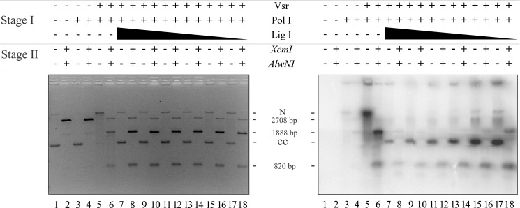
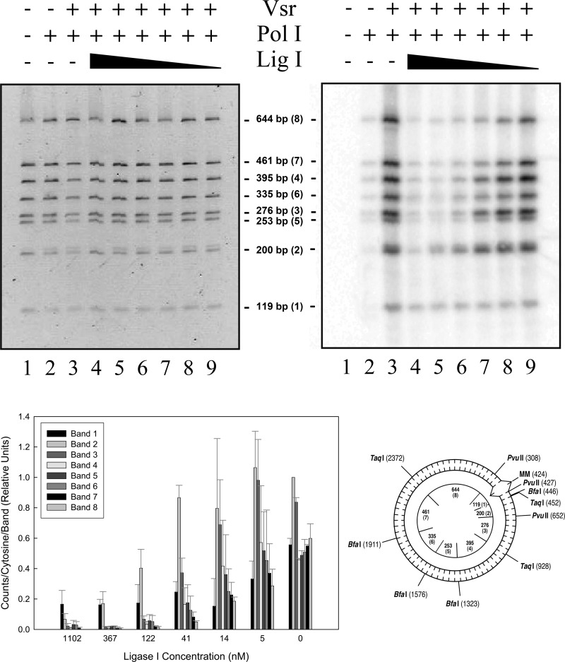
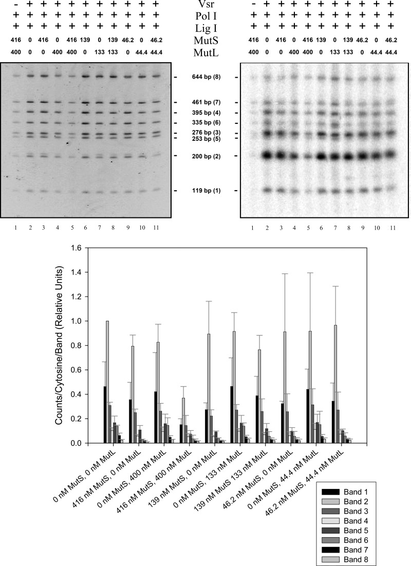
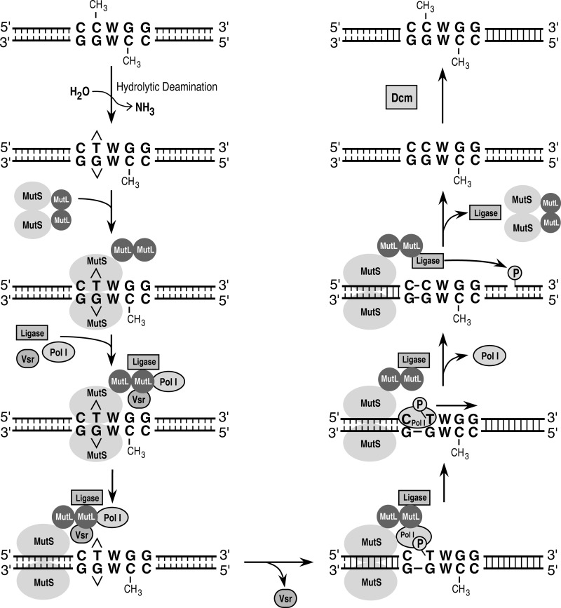
The Escherichia coli very short patch (VSP) repair pathway corrects thymidine-guanine mismatches that result from spontaneous hydrolytic deamination damage of 5-methyl cytosine. The VSP repair pathway requires the Vsr endonuclease, DNA polymerase I, a DNA ligase, MutS, and MutL to function at peak efficiency. The biochemical roles of most of these proteins in the VSP repair pathway have been studied extensively. However, these proteins have not been studied together in the context of VSP repair in an in vitro system. Using purified components of the VSP repair system in a reconstitution reaction, we have begun to develop an understanding of the role played by each of these proteins in the VSP repair pathway and have gained insights into their interactions. In this report we demonstrate an in vitro reconstitution of the VSP repair pathway using a plasmid DNA substrate. Surprisingly, the repair track length can be modulated by the concentration of DNA ligase. We propose roles for MutL and MutS in coordination of this repair pathway.


Figures

FIGURE 1.
FIGURE 1.
SDS-PAGE analysis of purified DNA polymerase I, DNA ligase I, and the Vsr endonuclease. Purified proteins (3 μg) were resolved on an 11% polyacrylamide gel run in the presence of SDS and stained with Coomassie Blue. Lane 1, molecular mass standards (size in kDa indicated on the left); lane 2, DNA polymerase I; lane 3, DNA ligase I; lane 4, the Vsr endonuclease.
FIGURE 2.
FIGURE 2.
Vsr-catalyzed nicking of a covalently closed circular DNA substrate. Vsr-dependent nicking reactions were as described under “Materials and Methods.” A, the titration of the Vsr endonuclease was from 153 to 0 nm with each DNA substrate, and incubation was for 10 min at 37 °C. The reactions were stopped, and the products were resolved on a 1.4% agarose gel containing 0.5 μg/ml ethidium bromide. The positions of nicked (N) and covalently closed (CC) DNA are noted on the right. A representative gel is shown. B, quantification of Vsr nicking reaction using covalently closed DNA substrates. The fraction of nicked DNA was determined as indicated under “Materials and Methods.” The data represent the average of at least three experiments with error bars indicating the standard deviation about the mean. The plasmids used were covalently closed DNA containing a T:G mismatch the Vsr recognition sequence (●), covalently closed homoduplex DNA (■), and covalently closed DNA containing a T:G mismatch outside the Vsr recognition sequence (▴).
FIGURE 3.
FIGURE 3.
The Vsr endonuclease incises DNA immediately 5′ to the mismatched thymidine. Vsr nicking reactions were as described under “Materials and Methods.” The Vsr endonuclease was incubated with covalently closed pUC19-VSR heteroduplex [32P]DNA (shown on the right; asterisk denotes location of 32P, and carats denote the G:T mismatch) for 10 min at 37 °C. The reactions were stopped, and the Vsr endonuclease was removed by organic extraction followed by EtOH precipitation. The circular DNA was incubated with EcoRI and BamHI, which flank the Vsr endonuclease recognition sequence as noted in the depiction of the substrate on the right. The resulting products were resolved on 16% denaturing polyacrylamide gel. Lane 1, no protein control; lane 2, 153 nm Vsr; lane 3, 51 nm Vsr; lane 4, 17 nm Vsr; lane 5, 5.7 nm Vsr; lane 6, 1.9 nm Vsr; lane 7, 0.6 nm Vsr.
FIGURE 4.
FIGURE 4.
DNA polymerase I and the Vsr endonuclease are sufficient to repair a T:G mismatch. The VSP repair reaction contained 50 ng of covalently closed heteroduplex substrate, 52 nm Vsr endonuclease, and a titration of DNA polymerase I from 467 to 0.2 nm as described under “Materials and Methods.” After incubation at 37 °C for 60 min, the reactions were stopped, DNA polymerase I and the Vsr endonuclease were removed by organic extraction, and the DNA was precipitated with EtOH. A, the DNA was digested with XcmI and AlwNI, and products were resolved on a 1.4% agarose gel containing 0.5 μg/ml ethidium bromide. Marker lanes are shown on both sides of the gel. B, experiments identical to the one shown in A were quantified as described under “Materials and Methods.” The data presented represent the averages of at least three experiments with error bars representing standard deviations from the mean. The fraction of DNA repaired was calculated as described under “Materials and Methods.”
FIGURE 5.
FIGURE 5.
DNA ligase I seals the nick created by DNA polymerase I nick translation. Repair and ligation reactions were conducted as described under “Materials and Methods” using 200 ng of pUC19-VSR heteroduplex DNA (∼1.2 nm molecules), 10 μm dATP, dGTP, or dTTP, and 0.5 μm [α-32P]dCTP, 52 nm Vsr endonuclease, 17 nm DNA polymerase I and a titration of DNA ligase I from 1100 to 5 nm. After incubation for 60 min at 37 °C, the reactions were stopped, the proteins were removed by organic extraction, and the DNA was precipitated with EtOH. One-quarter of each reaction was digested with XcmI and AlwNI to confirm that repair had occurred (even-numbered lanes), and another quarter of the reaction remained untreated (odd-numbered lanes). Both the digested and undigested reactions were run on a 1.4% agarose gel containing 0.5 μg/ml ethidium bromide. The left panel shows a gel stained with EtBr (0.5 μg/ml), confirming that repair had occurred. The right panel shows an autoradiogram demonstrating the incorporation of [α-32P]dCMP and ligation to form covalently closed circles after repair. The positions of nicked DNA (N), covalently closed DNA (CC), and the expected products of the restriction digest (2708, 1888, and 820 bp) are shown.
FIGURE 6.
FIGURE 6.
The impact of DNA ligase I concentration on repair track length. Ligation and repair reactions were as described under “Materials and Methods” using 200 ng of covalently closed pUC19-VSR heteroduplex DNA (∼1.2 nm molecules), 10 μm dATP, 10 μm dGTP, 10 μm dTTP, 0.5 μm [α-32P]dCTP, 26 μm NAD+, 50 nm Vsr endonuclease, 17 nm DNA polymerase I, and DNA ligase I (Lig I) at the indicated concentrations. After incubation for 60 min at 37 °C, the reactions were stopped, the proteins were removed by organic extraction, and the DNA was precipitated with EtOH. Each reaction was digested with BfaI, PvuII, and TaqI, and the products were resolved on a 6% native polyacrylamide gel followed by staining with 0.5 μg/ml ethidium bromide. The upper left panel shows the stained polyacrylamide gel, and the upper right panel shows the autoradiogram. The lower left panel shows quantification of [32P]dCMP incorporated into each band normalized to band 2 in the lane containing no DNA ligase I. The data presented represent the averages of at least two experiments with error bars representing standard deviations about the mean. The pUC19-VSR heteroduplex plasmid DNA substrate is shown in the lower right panel. The T:G mismatch within the canonical Vsr endonuclease recognition sequence is denoted as MM. Relevant restriction sites and their positions are shown on the outside circle. Fragments are listed by size with the relative distance from the mismatch shown in parentheses. Restriction fragment sizes generated by cleavage with BfaI, PvuII, and TaqI are shown inside the circle. The agarose gel run to demonstrate repair of the DNA substrate, similar to that shown in Fig. 5, is not shown.
FIGURE 7.
FIGURE 7.
MutS and MutL shorten VSP repair track lengths in vitro. Ligation and repair reactions were conducted as described in the legend for Fig. 5 with the addition of 1 mm ATP, a single concentration of DNA ligase I (4.5 nm), and the indicated concentration of MutS, MutL or both MutS and MutL. The reactions were processed and analyzed as described for Fig. 5. The upper left panel shows the stained polyacrylamide gel, and the upper right panel shows the autoradiogram. Fragments are listed by size with the relative distance from the mismatch shown in parentheses. The bottom panel shows quantification of [32P]dCMP incorporated into each band normalized to band 2 in the lane containing no MutS or MutL. The data presented represent the averages of at least two experiments with error bars representing standard deviations about the mean. The agarose gel run to demonstrate repair of the DNA substrate, similar to that shown in Fig. 5, is not shown.
FIGURE 8.
FIGURE 8.
A model for VSP repair. A T:G mismatch is generated by the spontaneous hydrolytic deamination of a 5-methyl cytosine residue. MutS and MutL are recruited to the T:G mismatched base, presumably through the interaction of MutS with the mismatch. MutS and MutL then recruit the Vsr endonuclease, DNA ligase I, and DNA polymerase I, effectively increasing the local concentration of each of the proteins. A physical interaction between MutL and the Vsr endonuclease has been demonstrated (24, 35); other protein-protein interactions, if they exist, are hypothetical. The Vsr endonuclease catalyzes the hydrolysis of a phosphodiester bond 5′ to the mismatched thymidine. DNA polymerase I is then loaded onto the nick and undergoes nick translates to repair the mismatch and synthesize a short repair patch. DNA ligase I seals the nick to restore the integrity of the DNA strand, and the Dcm methyltransferase methylates the appropriate cytosine on the newly synthesized repair patch.

Similar articles

Cited by

References

    1. Harfe B. D., Jinks-Robertson S. (2000) DNA mismatch repair and genetic instability. Annu. Rev. Genet. 34, 359–399 - PubMed
    1. Iyer R. R., Pluciennik A., Burdett V., Modrich P. L. (2006) DNA mismatch repair. Functions and mechanisms. Chem. Rev. 106, 302–323 - PubMed
    1. Jiricny J. (2006) The multifaceted mismatch-repair system. Nat. Rev. Mol. Cell Biol. 7, 335–346 - PubMed
    1. Schofield M. J., Hsieh P. (2003) DNA mismatch repair. Molecular mechanisms and biological function. Annu. Rev. Microbiol. 57, 579–608 - PubMed
    1. Kunkel T. A., Erie D. A. (2005) DNA mismatch repair. Annu. Rev. Biochem. 74, 681–710 - PubMed

Publication types

MeSH terms

LinkOut - more resources