. 2012 Sep 28;287(40):33812-25.
doi: 10.1074/jbc.M112.367839. Epub 2012 Jul 30.

Nuclear-to-cytoplasmic relocalization of the proliferating cell nuclear antigen (PCNA) during differentiation involves a chromosome region maintenance 1 (CRM1)-dependent export and is a prerequisite for PCNA antiapoptotic activity in mature neutrophils

Affiliations

Nuclear-to-cytoplasmic relocalization of the proliferating cell nuclear antigen (PCNA) during differentiation involves a chromosome region maintenance 1 (CRM1)-dependent export and is a prerequisite for PCNA antiapoptotic activity in mature neutrophils

Dikra Bouayad et al. J Biol Chem. .

Abstract

Neutrophils are deprived of proliferative capacity and have a tightly controlled lifespan to avoid their persistence at the site of injury. We have recently described that the proliferating cell nuclear antigen (PCNA), a nuclear factor involved in DNA replication and repair of proliferating cells, is a key regulator of neutrophil survival. In neutrophils, PCNA was localized exclusively in the cytoplasm due to its nuclear-to-cytoplasmic relocalization during granulocytic differentiation. We showed here that leptomycin B, an inhibitor of the chromosome region maintenance 1 (CRM1) exportin, inhibited PCNA relocalization during granulocytic differentiation of HL-60 and NB4 promyelocytic cell lines and of human CD34(+) primary cells. Using enhanced green fluorescent protein fusion constructs, we have demonstrated that PCNA relocalization involved a nuclear export signal (NES) located from Ile-11 to Ile-23 in the PCNA sequence. However, this NES, located at the inner face of the PCNA trimer, was not functional in wild-type PCNA, but instead, was fully active and leptomycin B-sensitive in the monomeric PCNAY114A mutant. To test whether a defect in PCNA cytoplasmic relocalization would affect its antiapoptotic activity in mature neutrophils, a chimeric PCNA fused with the SV40 nuclear localization sequence (NLS) was generated to preclude its cytoplasmic localization. As expected, neutrophil-differentiated PLB985 cells expressing ectopic SV40NLS-PCNA had an increased nuclear PCNA as compared with cells expressing wild-type PCNA. Accordingly, the nuclear PCNA mutant did not show any antiapoptotic activity as compared with wild-type PCNA. Nuclear-to-cytoplasmic relocalization that occurred during myeloid differentiation is essential for PCNA antiapoptotic activity in mature neutrophils and is dependent on the newly identified monomerization-dependent PCNA NES.


Figures
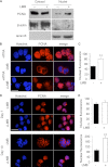
FIGURE 1.
FIGURE 1.
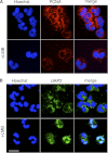
PCNA nuclear-to-cytoplasmic relocalization during granulocytic differentiation. A, effect of LMB on PCNA in nucleus and cytosol in ATRA-differentiated NB4 cells. Four days after ATRA treatment, NB4 cells were treated with solvent (−) or with 20 ng/ml LMB (+) for 4 h, and PCNA protein was analyzed by Western blot in nuclear and cytosolic fractions (10 and 20 μg/well, respectively). β-Actin and lamin B were used as specific marker for cytosol and nuclei and as loading controls, respectively. This representative experiment has been performed three times with identical results. B, effect of LMB on PCNA relocalization in HL60 cells. Four days after ATRA treatment, HL60 cells were treated with solvent (−) or with 20 ng/ml LMB (+) for 4 h, and PCNA localization was analyzed with confocal microscopy after immunolabeling using the Ab5 rabbit polyclonal anti-PCNA antibody. The nuclei were visualized with Hoechst staining (original magnification ×630, scale bar = 20 μm). This representative experiment has been performed four times with identical results. C, quantification of nuclear fluorescence intensity with the ImageJ 1.42 software in HL60 cells after LMB treatment. Quantification of nuclear PCNA is expressed in arbitrary units, and the means ± S.E. of four independent experiments in which more than 100 cells in each condition have been counted are given (**, p < 0.01, Student's t test). D, effect of LMB on PCNA relocalization during granulocytic differentiation of human CD34+ cells. CD34+/CD36 cells were treated with LMB (10 ng/ml or with solvent for 6 h) at day 7 (upper panel) or at day 13 (lower panel) after starting granulocytic differentiation with G-CSF/SCF/IL-3. PCNA localization was analyzed with confocal microscopy as in B. The nuclei were visualized with Hoechst staining (original magnification ×630, scale bar = 20 μm). This representative experiment has been performed three times with identical results. E, quantification of nuclear fluorescence intensity in CD34+/CD36 cells at day 7 (upper panel) or at day 13 (lower panel) in the presence or absence of LMB as in C. Data are expressed in arbitrary units, and the means ± S.E. of three independent experiments are given (**, p < 0.01, Student's t test).
FIGURE 2.
FIGURE 2.
Effect of LMB on PCNA and cIAP1 localization in mature neutrophils. Neutrophils were treated with solvent (−) or with 10 ng/ml LMB (+) for 5 h, and PCNA (A) and cIAP1 (B) localization was analyzed with confocal microscopy after immunolabeling. The nuclei were visualized with Hoechst staining (original magnification ×630, scale bar = 20 μm).
FIGURE 3.
FIGURE 3.
Localization and functional characterization of PCNA NES in HeLa cells. A, effect of NES1 and NES2 fusion on EGFP localization using either EGFP-N1 (upper panels) or EGFP-C3 (lower panels) plasmids, allowing us to fuse NES sequence at the amino- or carboxyl-terminal part of EGFP, respectively. HeLa cells were transfected with pEGFP-N1, pEGFP-C3, pNES1-EGFP, pEGFP-NES1, pNES2-EFGP, or pEGFP-NES2, and EGFP was detected by confocal scanning microscopy (original magnification ×630, scale bar = 20 μm). B, effect of LMB on NES1-EGFP localization. HeLa cells transfected with pNES1-EGFP were treated with either ethanol solvent (−) or 20 ng/ml LMB for 6 h prior to fixation and permeabilization. HeLa cells transfected with EGFP-N1 were used as controls. Nuclei were stained with propidium iodide (original magnification ×400, scale bar = 20 μm, left panel). C, the percentage of cells showing either strong nuclear localization (white bars, N>C) or strong cytoplasmic localization (black bars, N<C) was determined by direct counting. Counting was performed on a minimum of 100 transfected cells per experiment. The data are the means ± S.E. of three independent experiments, **, p <0.001, ANOVA test. D, effect of leucine into alanine mutations within NES1 on the NES1-EGFP localization. HeLa cells were transfected with pEGFP-N1, pNES1-EGFP, or pMutNES1-EGFP, and EGFP was detected with confocal scanning microscopy. Nuclei were visualized with Hoechst stain (original magnification ×400, scale bar = 20 μm, left panel). E, the percentages of cells showing nuclear and cytoplasmic EGFP localization were determined as in C. The data are the means ± S.E. of three independent experiments, **, p <0.001, ANOVA test.
FIGURE 4.
FIGURE 4.
Effect of NES1 mutation on PCNA subcellular localization. A, analysis of PCNA localization by indirect immunofluorescence using mouse monoclonal anti-HA antibody in HeLa cells transfected with pcDNA3-HA-PCNA constructs containing different mutations or deletions within the PCNA NES1. The cells were visualized using epifluorescence microscopy (original magnification ×400, scale bar = 20 μm). B, the percentages of cells showing strong nuclear PCNA localization (white bars, N>C) and pancellular localization (gray bars, N=C) are plotted in histograms. Counting was performed on a minimum of 100 transfected cells per experiment in three independent experiments. The data are the means ± S.E. of the three independent experiments, ***, p <0.001 (ANOVA test).
FIGURE 5.
FIGURE 5.
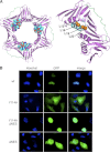
Nuclear localization of NES-deleted monomeric PCNA. A, tridimensional structure of trimeric and monomeric PCNA. Left panel: NES1 localization within the PCNA tridimensional structure (Protein Data Bank (PDB) ID: 1VYM). The interdomain connecting loop of PCNA is shown in green. The NES1 sequence is localized in the inner face of the PCNA trimer (cyan central helix). The protein is represented with graphics, and the NES1 atoms (Ile-11–Asn-23) are represented with balls colored following their type (nitrogen, blue; oxygen, red; carbon, cyan). Right panel: the Y114A mutation results in PCNA monomerization. The close-up of the mutated amino acids (orange balls) of the NES1 shows the following residues: Leu-12, Leu-16, Leu-19, and Leu-22. Both figures have been generated with PyMOL (42). B, analysis of GFP-PCNA localization by confocal scanning microscopy in HeLa cells transfected with pcDNA3-GFP-wtPCNA (wild-type PCNA), pcDNA3-GFP-PCNAY114A, pcDNA3-GFP-PCNAY114AΔ12–16NES1, or pcDNA3-GFP-PCNAΔ12–16NES1). Nuclei were visualized by Hoechst staining (original magnification ×630, scale bar = 20 μm).
FIGURE 6.
FIGURE 6.
NES deletion in monomeric PCNA NES is functional in monomeric PCNA. HeLa cells were transfected with pcDNA3-GFP-wtPCNA (WT PCNA) or pcDNA3-GFP-PCNAY114A (PCNAY114A). A, effect of LMB on the monomeric PCNA mutant PCNAY114A. HeLa cells expressing PCNAY114A were treated with either ethanol solvent (−) or 20 ng/ml LMB for 6 h prior to fixation and permeabilization, and cells were analyzed by scanning confocal microscopy. HeLa cells transfected with pcDNA3-GFP-wtPCNA and treated with solvent were used as controls. B, colocalization of CRM1 with PCNAY114A but not with WT PCNA in HeLa cells. Immunofluorescence detection of CRM1 was performed using a rabbit polyclonal anti-CRM1 antibody followed by an Alexa Fluor 555-coupled goat anti-rabbit IgG, and cells were analyzed by scanning confocal microscopy. A and B show representative experiments that have been performed at least three times with similar results. The nuclei were visualized with Hoechst staining (original magnification ×630, scale bar = 20 μm).
FIGURE 7.
FIGURE 7.
Localization of the SV40NLS-PCNA mutant in PLB985 cells before and after DMF-induced granulocytic differentiation. A and B, immunofluorescence analysis of PCNA localization in PLB985 cells transfected with pcDNA3-PCNA (wild-type WT PCNA) and pcDNA3-SV40NLS-PCNA before (− DMF) (A) and after (+ DMF) (B) granulocytic differentiation. Immunofluorescence analysis of PCNA was performed with the Ab5 rabbit polyclonal anti-PCNA antibody, and the cells were analyzed with scanning confocal microscopy. The nuclei were visualized with Hoechst staining (scale bar = 20 μm). This representative experiment has been performed at least three times with similar results. C, quantification of nuclear fluorescence intensity with the ImageJ 1.42 software. WT-PCNA- and SV40NLS-PCNA-transfected PLB985 cells are represented by black and gray bars, respectively. Fluorescence intensities have been determined in more than 100 cells in each of the four conditions and were expressed in arbitrary units. Data are means ± S.E. (**, p < 0.01, ANOVA test). AU, arbitrary units. D, Western blot analysis of PCNA protein in nuclear (20 μg/well) and cytosolic (10 μg/well) fractions. β-Actin and lamin B were used as specific marker for cytosol and nuclei and as loading control, respectively.
FIGURE 8.
FIGURE 8.
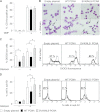
Defect in antiapoptotic activity of the nuclear SV40NLS-PCNA mutant in neutrophil-differentiated PLB985 cells. A, CD11b membrane expression in neutrophil-differentiated PLB985 cells stably overexpressing wild-type PCNA or the nuclear SV40NLS-PCNA mutant as compared with control PLB985 cells (empty plasmid). PLB985 cells were treated with DMF for 5 days to induce granulocyte differentiation. CD11b expression was measured by flow cytometry and expressed as the percentage of CD11b-positive cells. B–D, neutrophil-differentiated PLB985 cells stably overexpressing wild-type PCNA or the nuclear SV40NLS-PCNA mutant or the control PLB985 cells were incubated with or without 1 μg/ml gliotoxin for 16 h to induce apoptosis. B, May Grunwald Giemsa staining showing cells with condensed nuclei indicative of an apoptotic morphology (shown by black arrows) in gliotoxin-treated cells (lower panels) as compared with untreated cells (upper panels). The typical experiment shown in B was representative of four independent experiments. The percentages of cells with an apoptotic morphology after gliotoxin treatment were 41.4% ± 8, 22.5% ± 7, and 46.9% ± 10 in PLB985-Empty plasmid, PLB985-PCNA, and PLB985-SV40NLS-PCNA, respectively. Data are means ± S.E. (more than 100 cells in each condition have been counted for each experiment). C, mitochondrial depolarization analysis after DIOC6 labeling in the neutrophil-differentiated PLB985 cells. The histogram in the left panel represents the percentage of apoptotic cells with decreased mitochondrial potential, and data are expressed as the means ± S.E. of nine independent experiments (*, p < 0.05, ANOVA test). Representative flow cytometry plots showing the percentage of cells with depolarized mitochondria with decreased DIOC6 labeling are depicted in the right panel. D, the percentage of neutrophil-differentiated PLB985 cells in the sub-G1 phase showing DNA fragmentation after propidium iodide staining. The histogram on the left panel depicts the means ± S.E. of six independent experiments (*, p < 0.05, ANOVA test), and representative flow cytometry plots are shown in the right panel.

Similar articles

Cited by

References

    1. Borregaard N. (2010) Neutrophils, from marrow to microbes. Immunity 33, 657–670 - PubMed
    1. Witko-Sarsat V., Rieu P., Descamps-Latscha B., Lesavre P., Halbwachs-Mecarelli L. (2000) Neutrophils: molecules, functions, and pathophysiological aspects. Lab. Invest. 80, 617–653 - PubMed
    1. Simon H. U. (2003) Neutrophil apoptosis pathways and their modifications in inflammation. Immunol. Rev. 193, 101–110 - PubMed
    1. Fox S., Leitch A. E., Duffin R., Haslett C., Rossi A. G. (2010) Neutrophil apoptosis: relevance to the innate immune response and inflammatory disease. J. Innate Immun. 2, 216–227 - PMC - PubMed
    1. Rossi A. G., Sawatzky D. A., Walker A., Ward C., Sheldrake T. A., Riley N. A., Caldicott A., Martinez-Losa M., Walker T. R., Duffin R., Gray M., Crescenzi E., Martin M. C., Brady H. J., Savill J. S., Dransfield I., Haslett C. (2006) Cyclin-dependent kinase inhibitors enhance the resolution of inflammation by promoting inflammatory cell apoptosis. Nat. Med. 12, 1056–1064 - PubMed

Publication types

MeSH terms