

Ocular ischemic syndrome - a systematic review
- PMID: 22847215
- PMCID: PMC3560693
- DOI: 10.12659/msm.883260
Ocular ischemic syndrome - a systematic review
Abstract
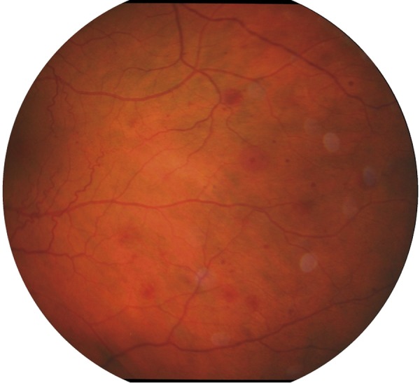
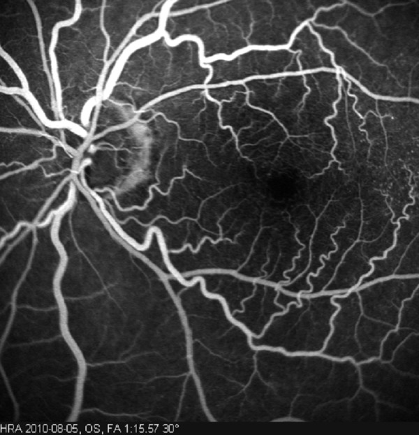
Ocular ischemic syndrome is a rare condition, which is caused by ocular hypoperfusion due to stenosis or occlusion of the common or internal carotid arteries. Atherosclerosis is the major cause of changes in the carotid arteries. Ocular ischemic syndrome is manifested as visual loss, orbital pain and, frequently, changes of the visual field, and various anterior and posterior segment signs. Anterior segment signs include iris neovascularization and secondary neovascular glaucoma, iridocyclitis, asymmetric cataract, iris atrophy and sluggish reaction to light. Posterior eye segment changes are the most characteristic, such as narrowed retinal arteries, perifoveal telangiectasias, dilated retinal veins, mid-peripheral retinal hemorrhages, microaneurysms, neovascularization at the optic disk and in the retina, a cherry-red spot, cotton-wool spots, vitreous hemorrhage and normal-tension glaucoma. Differential diagnosis of ocular ischemic syndrome includes diabetic retinopathy and moderate central retinal vein occlusion. Carotid artery imaging and fundus fluorescein angiography help to establish the diagnosis of ocular ischemic syndrome. The treatment can be local, for example, ocular (conservative, laser and surgical) or systemic (conservative and surgical treatment of the carotid artery). Since the condition does not affect the eyes alone, patients with ocular ischemic syndrome should be referred for consultation to the neurologist, vascular surgeon and cardiologist.
Figures
References
-
- Hedges TR., Jr Ophthalmoscopic findings in internal carotid artery occlusion. Am J Ophthalmol. 1963;55:1007–12. - PubMed
-
- Kearns TP, Hollenhorst RW. Venous stasis retinopathy of occlusive disease of the carotid artery. Mayo Clin Proc. 1963;38:304–12. - PubMed
-
- Mendrinos E, Machinie TG, Pournaras CJ. Ocular Ischemic Syndrome. Surv Ophthalmol. 2010;55(1):2–34. - PubMed
-
- Sharma S, Brown GC. In: Ocular Ischemic Syndrome. Ryan SJ, Hinton DR, Schachat AP, et al., editors. Elsevier; 2006. pp. 1491–502.
-
- Brown GC, Magargal LE. The ocular ischemic syndrome. Clinical, fluorescein angiographic and carotid angiographic features. Int Ophthalmol. 1988;11(4):239–51. - PubMed
Publication types
MeSH terms
LinkOut - more resources
Full Text Sources
Other Literature Sources
Medical
