. 2012 Aug 14;109(33):13278-83.
doi: 10.1073/pnas.1203536109. Epub 2012 Jul 30.

Structure of the rhesus monkey TRIM5α PRYSPRY domain, the HIV capsid recognition module

Affiliations

Structure of the rhesus monkey TRIM5α PRYSPRY domain, the HIV capsid recognition module

Nikolaos Biris et al. Proc Natl Acad Sci U S A. .

Abstract

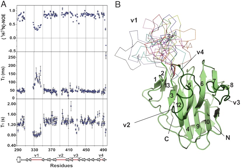
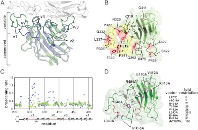
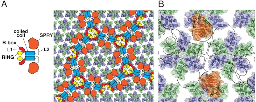
Tripartite motif protein TRIM5α blocks retroviral replication after cell entry, and species-specific differences in its activity are determined by sequence variations within the C-terminal B30.2/PRYSPRY domain. Here we report a high-resolution structure of a TRIM5α PRYSPRY domain, the PRYSPRY of the rhesus monkey TRIM5α that potently restricts HIV infection, and identify features involved in its interaction with the HIV capsid. The extensive capsid-binding interface maps on the structurally divergent face of the protein formed by hypervariable loop segments, confirming that TRIM5α evolution is largely determined by its binding specificity. Interactions with the capsid are mediated by flexible variable loops via a mechanism that parallels antigen recognition by IgM antibodies, a similarity that may help explain some of the unusual functional properties of TRIM5α. Distinctive features of this pathogen-recognition interface, such as structural plasticity conferred by the mobile v1 segment and interaction with multiple epitopes, may allow restriction of divergent retroviruses and increase resistance to capsid mutations.


Conflict of interest statement

The authors declare no conflict of interest.

Figures

Fig. 1.
Fig. 1.
PRYSPRY structure and dynamics. (A) The 15N T1, T2 and {15N-1H} NOE measurements of the rhTRIM5α PRYSPRY. (B), The structure of the rhTRIM5α PRYSPRY domain. Ten lowest-energy conformations of the v1 loop calculated using NOE restraints are shown as colored ribbons. The rest of the structure determined by X-ray crystallography is shown as a light green cartoon. The strands forming the beta sandwich PRYSPRY fold are numbered, and the variable loop regions are labeled and highlighted in dark green.
Fig. 2.
Fig. 2.
The capsid-binding surface of PRYSPRY. (A) rhTRIM5α PRYSPRY (green) superimposed on the structure of TRIM21 PRYSPRY (blue) in complex with the IgG Fc chain (white). Structurally divergent variable loops (v1 through v4) are highlighted and labeled. (B) The view of the predicted capsid-interacting surface of the rhTRIM5α PRYSPRY. Residues under positive evolutionary selection are colored red and labeled (13). The mobile surface formed by the v1 loop is colored yellow, whereas v2, v3, and v4 segments are highlighted in dark green. (C) The broadening rates of the 15N TROSY HSQC signals plotted for all assigned residues of the wild-type rhTRIM5α PRYSPRY (blue) and the v1C-5A PRYSPRY mutant (green). (D) The view of the capsid-binding surface illustrating the results of NMR titrations (Fig. 2C) and mutagenesis. Backbone segments perturbed by HIV CA-NTD titrations are colored in red, unaffected in green, and unassigned in gray. Residues tested by point mutagenesis are shown in magenta, whereas the segment changed to five alanines in the v1C-5A mutant is shown in cyan. HIV-1 restriction activity measurements for mutations located on the variable face of the rhTRIM5α PRYSPRY are listed in the table.
Fig. 3.
Fig. 3.
Possible arrangement of the TRIM5α PRYSPRY on the surface of the HIV-1 mature core. (A) Domain structure of TRIM5α and one of the several possible relative orientations of the hexagonal arrays formed by TRIM5α and the HIV capsid (adapted from ref. (15)). HIV capsid assembly is derived from PDB ID codes 3DIK (47) and 3H47 (48). Only the N-terminal domains of the capsid that form the outer surface of the core are shown. Capsid C-terminal domains are omitted for clarity because they face the interior and are not accessible to interact with the PRYSPRY. Capsid monomers within the hexamers are shown in alternating colors (green and blue). (B) The structure of the rhTRIM5α PRYSPRY (orange) with the variable interaction face oriented toward the surface of the assembled capsid. The figure is only meant to illustrate the relative sizes of the PRYSPRY, CA-NTD, and the hexagonal arrays formed by the TRIM5α and the HIV capsid.

References

    1. Stremlau M, et al. The cytoplasmic body component TRIM5alpha restricts HIV-1 infection in Old World monkeys. Nature. 2004;427:848–853. - PubMed
    1. Ozato K, Shin DM, Chang TH, Morse HC., III TRIM family proteins and their emerging roles in innate immunity. Nat Rev Immunol. 2008;8:849–860. - PMC - PubMed
    1. McNab FW, Rajsbaum R, Stoye JP, O’Garra A. Tripartite-motif proteins and innate immune regulation. Curr Opin Immunol. 2011;23:46–56. - PubMed
    1. Pertel T, et al. TRIM5 is an innate immune sensor for the retrovirus capsid lattice. Nature. 2011;472:361–365. - PMC - PubMed
    1. Han K, Lou DI, Sawyer SL. Identification of a genomic reservoir for new TRIM genes in primate genomes. PLoS Genet. 2011;7:e1002388. - PMC - PubMed

Publication types

Substances

LinkOut - more resources