

Carcinosarcoma ex non-recurrent pleomorphic adenoma composed of TTF-1 positive large cell neuroendocrine carcinoma and myofibrosarcoma: apropos a rare Case
- PMID: 22847723
- PMCID: PMC3642266
- DOI: 10.1007/s12105-012-0385-0
Carcinosarcoma ex non-recurrent pleomorphic adenoma composed of TTF-1 positive large cell neuroendocrine carcinoma and myofibrosarcoma: apropos a rare Case
Abstract
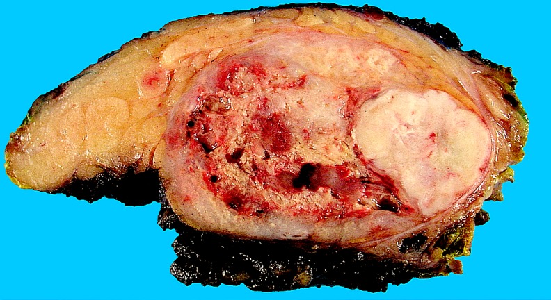
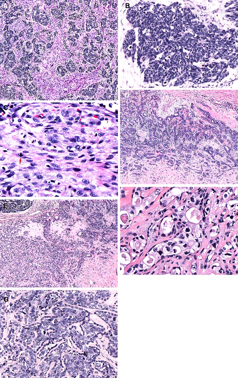
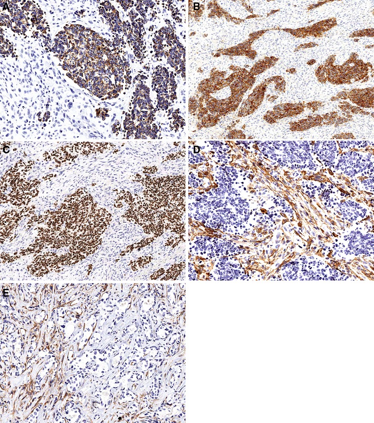
We present a carcinosarcoma ex non-recurrent pleomorphic adenoma composed of a large cell neuroendocrine carcinomatous component and a spindle cell sarcoma with myofibroblastic differentiation. The tumor contained a hyalinized transition zone where the classical PA appeared to acquire two different histopathological patterns of malignant transformation of the epithelial component. The carcinomatous component was strongly and diffusely positive for low-molecular weight cytokeratins (AE1-3), synaptophysin, thyroid transcription factor-1 and focally positive for chromogranin A. All these markers were negative in the sarcomatous component. The sarcomatous component displayed immunoreactivity for smooth muscle actin with a predominantly linear, subplasmalemmal pattern. No expression of CD31, S100 protein, h-caldesmon, desmin, CD34, p63, myogenin, Myo D1 and c-kit was detected. Strong immunohistochemical expression of p53 was documented in both the carcinomatous and sarcomatous components as well as in the atypical epithelial component in the transition zone associated with the hyalinized pleomorphic adenoma.
Figures
Similar articles
-
Carcinosarcoma ex Pleomorphic Adenoma of the Submandibular Gland in a 64-Year-Old Man: A Case Report.J Nippon Med Sch. 2018;85(1):51-55. doi: 10.1272/jnms.2018_85-8. J Nippon Med Sch. 2018. PMID: 29540647
-
Pulmonary carcinomas with pleomorphic, sarcomatoid, or sarcomatous elements: a clinicopathologic and immunohistochemical study of 75 cases.Am J Surg Pathol. 2003 Mar;27(3):311-24. doi: 10.1097/00000478-200303000-00004. Am J Surg Pathol. 2003. PMID: 12604887
-
Large cell neuroendocrine carcinoma with sarcomatous changes of the endometrium: a case report with immunohistochemical studies and molecular genetic study of KIT and PDGFRA.Pathol Res Pract. 2010 Jun 15;206(6):420-5. doi: 10.1016/j.prp.2009.12.008. Epub 2010 Feb 26. Pathol Res Pract. 2010. PMID: 20189318
-
Minimally invasive carcinosarcoma ex pleomorphic adenoma: A case report and literature review with cytohistological correlation.Head Neck. 2016 Sep;38(9):E2483-9. doi: 10.1002/hed.24462. Epub 2016 Apr 15. Head Neck. 2016. PMID: 27080524 Review.
-
Myofibrosarcoma.Virchows Arch. 2004 Sep;445(3):215-23. doi: 10.1007/s00428-004-1038-9. Epub 2004 Jun 2. Virchows Arch. 2004. PMID: 15173943 Review.
Cited by
-
Large cell neuroendocrine carcinoma of the nasal cavity: an extremely rare and new distinct entity.Pan Afr Med J. 2018 Jun 29;30:188. doi: 10.11604/pamj.2018.30.188.14992. eCollection 2018. Pan Afr Med J. 2018. PMID: 30455817 Free PMC article.
-
Intraparotid Ganglioneuroma: A rare case report.Indian J Otolaryngol Head Neck Surg. 2023 Sep;75(3):2613-2616. doi: 10.1007/s12070-023-03800-7. Epub 2023 May 8. Indian J Otolaryngol Head Neck Surg. 2023. PMID: 37636636 Free PMC article.
-
Large Cell Neuroendocrine Carcinoma of the Head and Neck: A Clinicopathologic Series of 10 Cases With an Emphasis on HPV Status.Am J Surg Pathol. 2016 Apr;40(4):471-8. doi: 10.1097/PAS.0000000000000580. Am J Surg Pathol. 2016. PMID: 26735857 Free PMC article.
-
Clinical and Morphological Aspects of Aggressive Salivary Gland Mixed Tumors: A Narrative Review.Diagnostics (Basel). 2024 Sep 3;14(17):1942. doi: 10.3390/diagnostics14171942. Diagnostics (Basel). 2024. PMID: 39272728 Free PMC article. Review.
-
A Case of a Coexisting Carcinosarcoma Ex Pleomorphic Adenoma With Langerhans Cell Histiocytosis in the Parotid Gland.Cureus. 2023 Jul 24;15(7):e42351. doi: 10.7759/cureus.42351. eCollection 2023 Jul. Cureus. 2023. PMID: 37621779 Free PMC article.
References
-
- Gnepp DR. Malignant mixed tumors of the salivary glands: a review. Pathol Annu. 1993;28(Pt 1):279–328. - PubMed
-
- Gnepp DR. WHO classification of tumors. In: Barnes L, Eveson JW, Reichart P, Sidransky D, editors. Pathology and genetics. Head and neck tumors. Lyon: IARC Press; 2005.
-
- Kirklin JW, Mc DJ, Harrington SW, et al. Parotid tumors: histopathology, clinical behavior, and end results. Surg Gynecol Obstet. 1951;92:721–733. - PubMed
-
- Weiler C, Zengel P, van der Wal JE, et al. Carcinoma ex pleomorphic adenoma with special reference to the prognostic significance of histological progression: a clinicopathological investigation of 41 cases. Histopathology 2011;59:741–50. - PubMed
Publication types
MeSH terms
Substances
LinkOut - more resources
Full Text Sources
Research Materials
Miscellaneous
