. 2013 Jan;101(1):203-12.
doi: 10.1002/jbm.a.34309. Epub 2012 Jul 30.

Correlating macrophage morphology and cytokine production resulting from biomaterial contact

Affiliations

Correlating macrophage morphology and cytokine production resulting from biomaterial contact

Hyun-Su Lee et al. J Biomed Mater Res A. 2013 Jan.

Abstract

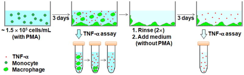
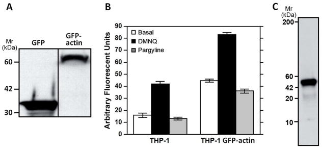
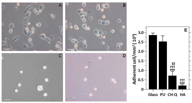
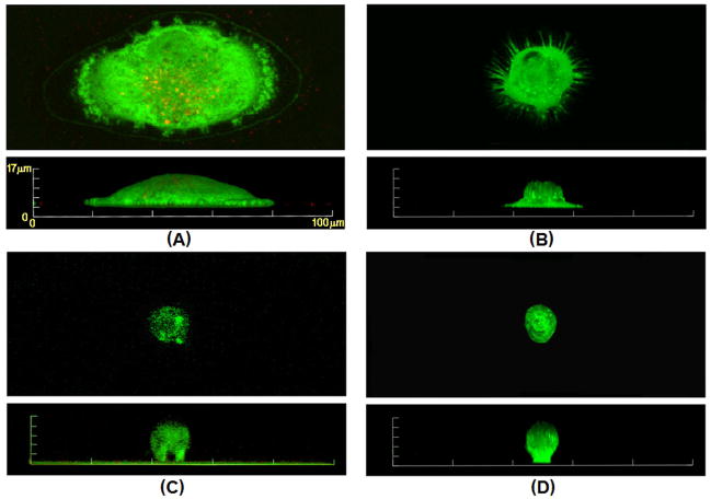
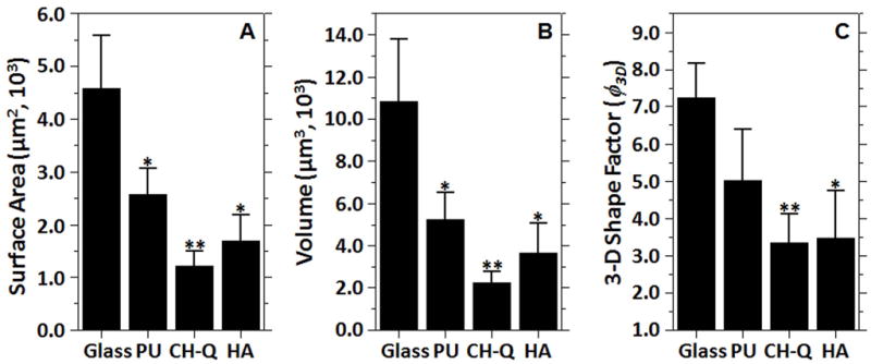
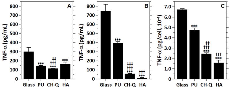
The morphological and inflammatory responses of adherent macrophages are correlated to evaluate the biocompatibility of surfaces. Monocyte-derived macrophage (MDM), THP-1, and THP-1 cells expressing GFP-actin chimeric protein were seeded onto glass, polyurethane (PU), and glass surface modified with quaternary ammonium salt functionalized chitosan (CH-Q) and hyaluronic acid (HA). Using confocal microscopy, the surface area, volume and 3D shape factor of adherent macrophages was quantified. For comparison, functional consequences of cell-surface interactions that activate macrophages and thereby elicit secretion of a proinflammatory cytokine were evaluated. Using an enzyme linked immune sorbent assay, tumor necrosis factor-alpha (TNF-α) was measured. On glass, macrophages exhibited mainly an amoeboid shape, exhibited the largest surface area, volume, and 3D shape factor and produced the most TNF-α. On PU, macrophages displayed mainly a hemispherical shape, exhibited an intermediate volume, surface area and 3D shape factor, and produced moderate TNF-α. In contrast, on CH-Q and HA surfaces, macrophages were spherical, exhibited the smallest volume, surface area, and 3D shape factor, and produced the least TNF-α. These studies begin to validate the use of GFP-actin-modified MDM as a novel tool to correlate cell morphology with inflammatory cell response.


Figures

FIGURE 1
FIGURE 1
Experimental scheme for measurement of TNF-α secretion by adherent macrophages (PMA-differentiated THP-1) on surfaces of glass, polyurethane (PU), chitosan possessing quaternary ammonium salts (CH-Q) and hyaluronic acid (HA). The cell culture medium volume is 7 mL for these experiments.
FIGURE 2
FIGURE 2
(A) THP-1 was transduced with the GFP-actin gene via a lentiviral vector (THP-1 GFP-actin). Western blot analysis of GFP expression from THP-GA lysates expressing GFP control or GFP-actin confirms the presence of the chimeric protein. (B) Cultured THP-1 cells (105) or THP-1 GFP-actin cells were seeded on PE films inserted on the bottom of 96 well plates. After 2 hr the culture medium was replaced with DHR-123 containing medium and cells were incubated for 2 hr at 37°C. Cells were rinsed twice with PBS, and 10−5 M of pargyline, a monoamine oxidase antagonist, or a superoxide initiator DMNQ was added to triplicate wells. Fluorescence (500 nm excitation, 536 nm emission) was measured at one hr. Higher values in the transduced cells are due to GFP expression. Results show identical trends in ROS production irrespective of GFP-actin treatment, thus strongly suggesting that the GFP-actin expression had no untoward effect upon normal MDM function. Data are presented as mean ± SD (n = 4 experiments). (C) Representative Western blot analysis of THP-1 GFP-actin expressing cell lysates immunoprecipitated with GFP-actin antibody and probed for the expression of the actin-binding protein Fascin.
FIGURE 3
FIGURE 3
Morphology of monocyte-derivatized macrophages (PMA-treated THP-1 cells) on (A) glass, (B) PU, (C) CH-Q, and (D) HA after three days. (A) Macrophages on the glass show amoeboid morphology, similar to macrophages adherent to polystyrene culture dishes. (B) Macrophages on PU demonstrate a round morphology. (C) Macrophages on CH-Q show a round morphology, a low cell surface density and small size, similar to cells attached to HA as shown in (D). Scale bar length is 50 μm. (E) Adhesion density of macrophages on the four surface types after three days. Data are presented as mean ∓standard deviation (n = 3 experiments). Statistical significance: ***P < 0.001 versus glass, †††P < 0.001 versus PU, ‡‡P < 0.01 versus HA, ‡‡‡P <0.001 versus HA.
FIGURE 4
FIGURE 4
Representative 3-D confocal fluorescence images of monocyte-derived macrophages (PMA-treated GFP-actin transduced THP-1 cells) on (A) glass, (B) PU, (C) CH-Q and (D) HA surfaces after three days of culturing. The images represent maximal cell projection along the optical axis (z-axis, top view in each panel A–D) and a side projection (y-axis, side view in each panel A–D). The identical y and z scales shown in (A) were used for all images.
FIGURE 5
FIGURE 5
(A) Cell surface area and (B) cell volume for macrophages adherent to glass, PU, CH-Q and HA surfaces. (C) Degree of cell spreading for macrophages adherent to the four surface types. The calculated 3-D shape factor φ3D = 1 for a perfectly spherical object. Data are presented as mean ±standard deviation (n = 3 experiments). Statistical significance: *P < 0.05 versus glass, **P < 0.01 versus glass.
FIGURE 6
FIGURE 6
(A) ELISA assay results of TNF-αlevels secreted by suspended and adherent macrophages cultured in glass dishes and PU-coated, CH-Q coated, HA coated glass dishes for three days. (B) TNF-αsecretion from adherent macrophages cultured for three additional days in each dish after rinsing and replacing the medium to remove suspended cells. (C) Normalized TNF-αsecretion levels per adherent cell on each surface type. Data are presented as mean ±standard deviation (n = 3 experiments). Statistical significance: ***P < 0.001 versus glass, †††P < 0.001 versus PU, ‡‡P < 0.01 versus HA, ‡‡‡P < 0.001 versus HA.

Similar articles

Cited by

References

    1. Richards MJ, Edwards JR, Culver DH, Gaynes RP. Nosocomial infections in medical intensive care units in the United States. Crit Care Med. 1999;27:887–892. - PubMed
    1. Markowicz P, Wolff M, Djedaini K, Cohen Y, Chastre J, Delclaux C, Merrer J, Herman B, Veber B, Fontaine A, Dreyfuss D. Multicenter prospective study of ventilator-associated pneumonia during acute respiratory distress syndrome - Incidence, prognosis, and risk factors. Am J Resp Crit Care. 2000;161:1942–1948. - PubMed
    1. Martin DC, O’Ryan FS, Indresano AT, Bogdanos P, Wang B, Hui RL, Lo JC. Characteristics of implant failures in patients with a history of oral bisphosphonate therapy. J Oral Maxil Surg. 2010;68:508–514. - PubMed
    1. Baddour LM, Bettmann MA, Bolger AF, Epstein AE, Ferrieri P, Gerber MA, Gewitz MH, Jacobs AK, Levison ME, Newburger JW, Pallasch TJ, Wilson WR, Baltimore RS, Falace DA, Shulman ST, Tani LY, Taubert KA. Nonvalvular cardiovascular device-related infections. Circulation. 2003;108:2015–2031. - PubMed
    1. Bernacca GM, Mackay TG, Wilkinson R, Wheatley DJ. Calcification and fatigue failure in a polyurethane heart valve. Biomaterials. 1995;16:279–285. - PubMed

Publication types

MeSH terms

LinkOut - more resources