

Effects of oxygen-glucose deprivation on microglial mobility and viability in developing mouse hippocampal tissues
- PMID: 22847985
- PMCID: PMC3786781
- DOI: 10.1002/glia.22394
Effects of oxygen-glucose deprivation on microglial mobility and viability in developing mouse hippocampal tissues
Abstract
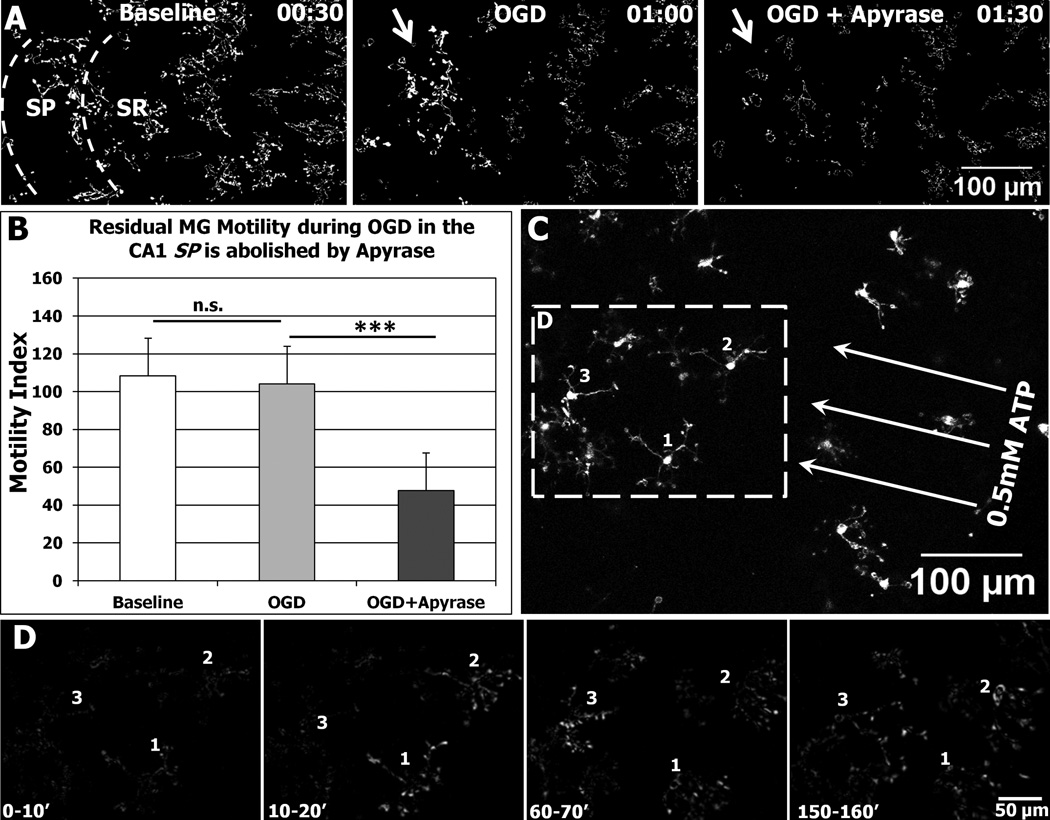
As brain-resident immune cells, microglia (MG) survey the brain parenchyma to maintain homeostasis during development and following injury. Research in perinatal stroke, a leading cause of lifelong disability, has implicated MG as targets for therapeutic intervention during stroke. Although MG responses are complex, work in developing rodents suggests that MG limit brain damage after stroke. However, little is known about how energy-limiting conditions affect MG survival and mobility (motility and migration) in developing brain tissues. Here, we used confocal time-lapse imaging to monitor MG viability and mobility during hypoxia or oxygen-glucose deprivation (OGD) in hippocampal tissue slices derived from neonatal GFP-reporter mice (CX3CR1(GFP/+) ). We found that MG remain viable for at least 6 h of hypoxia but begin to die after 2 h of OGD, while both hypoxia and OGD reduce MG motility. Unexpectedly, some MG retain or recover motility during OGD and can engulf dead cells. Additionally, MG from younger neonates (P2-P3) are more resistant to OGD than those from older ones (P6-P7), indicating increasing vulnerability with developmental age. Finally, transient (2 h) OGD also increases MG death, and although motility is rapidly restored after transient OGD, it remains below control levels for many hours. Together, these results show that MG in neonatal mouse brain tissues are vulnerable to both transient and sustained OGD, and many MG die within hours after onset of OGD. Preventing MG death may, therefore, provide a strategy for promoting tissue restoration after stroke.
Copyright © 2012 Wiley Periodicals, Inc.
Conflict of interest statement
The authors declare that they have no conflict of interest.
Figures


References
-
- Burguillos MA, Deierborg T, Kavanagh E, Persson A, Hajji N, Garcia-Quintanilla A, Cano J, Brundin P, Englund E, Venero JL, Joseph B. Caspase signalling controls microglia activation and neurotoxicity. Nature. 2011;472:319–324. - PubMed
-
- Carlsson Y, Schwendimann L, Vontell R, Rousset CI, Wang X, Lebon S, Charriaut-Marlangue C, Supramaniam V, Hagberg H, Gressens P, Jacotot E. Genetic inhibition of caspase-2 reduces hypoxic-ischemic and excitotoxic neonatal brain injury. Ann Neurol. 2011;70:781–789. - PubMed
-
- Cavaliere F, Dinkel K, Reymann K. Microglia response and P2 receptor participation in oxygen/glucose deprivation-induced cortical damage. Neuroscience. 2005;136:615–623. - PubMed
-
- Cavaliere F, Florenzano F, Amadio S, Fusco FR, Viscomi MT, D'Ambrosi N, Vacca F, Sancesario G, Bernardi G, Molinari M, Volonte C. Up-regulation of P2X2, P2X4 receptor and ischemic cell death: prevention by P2 antagonists. Neuroscience. 2003;120:85–98. - PubMed
-
- Chock VY, Giffard RG. Development of neonatal murine microglia in vitro: changes in response to lipopolysaccharide and ischemia-like injury. Pediatr Res. 2005;57:475–480. - PubMed
Publication types
MeSH terms
Substances
Grants and funding
LinkOut - more resources
Full Text Sources
Research Materials
