. 2012 Oct;40(19):9417-28.
doi: 10.1093/nar/gks723. Epub 2012 Jul 30.

DNA of a circular minichromosome linearized by restriction enzymes or other reagents is resistant to further cleavage: an influence of chromatin topology on the accessibility of DNA

Affiliations

DNA of a circular minichromosome linearized by restriction enzymes or other reagents is resistant to further cleavage: an influence of chromatin topology on the accessibility of DNA

Sławomir Kumala et al. Nucleic Acids Res. 2012 Oct.

Abstract

The accessibility of DNA in chromatin is an essential factor in regulating its activities. We studied the accessibility of the DNA in a ∼170 kb circular minichromosome to DNA-cleaving reagents using pulsed-field gel electrophoresis and fibre-fluorescence in situ hybridization on combed DNA molecules. Only one of several potential sites in the minichromosome DNA was accessible to restriction enzymes in permeabilized cells, and in growing cells only a single site at an essentially random position was cut by poisoned topoisomerase II, neocarzinostatin and γ-radiation, which have multiple potential cleavage sites; further sites were then inaccessible in the linearized minichromosomes. Sequential exposure to combinations of these reagents also resulted in cleavage at only a single site. Minichromosome DNA containing single-strand breaks created by a nicking endonuclease to relax any unconstrained superhelicity was also cut at only a single position by a restriction enzyme. Further sites became accessible after ≥95% of histones H2A, H2B and H1, and most non-histone proteins were extracted. These observations suggest that a global rearrangement of the three-dimensional packing and interactions of nucleosomes occurs when a circular minichromosome is linearized and results in its DNA becoming inaccessible to probes.


Figures
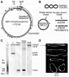
Figure 1.
Figure 1.
(A) The circular DNA of the EBV minichromosome. TR is the terminal repeated sequence through which linear EBV DNA is circularized to form the minichromosome, oriP is the preferred but not unique origin of replication, MAR is the nuclear matrix attachment region which coincides with a micrococcal nuclease-hypersensitive region. Approximate positions of regions transcribed in Raji cells are shown by arrows; black, highest level; white, intermediate; dashed, lowest (22). (B) Forms of minichromosome DNA considered in this work and (C) their migration in a PFGE gel of total cell DNA shown by hybridizing with an EBV DNA probe. In all gel images, the top coincides with the sample wells and each panel shows lanes from the same gel. Cells encapsulated in agarose beads and deproteinized were incubated with: C, no addition (2 h); PacI (100 U/ml, 2 h) which cuts minichromosome DNA at a single site; NbB, nicking endonuclease Nb.BbvCI (100 U/ml, 1 h). Lane virus DNA, linear EBV DNA; lane λ, oligomers of λ DNA (48.5 kb). (D) Representative DNA molecules extracted from the region close to the origin after incubating cells with Nb.BbvCI, stained with YOYO-1 and spread by molecular combing; these are believed to be nicked circular minichromosome DNA (see text).
Figure 2.
Figure 2.
Minichromosome DNA is cut at only one site by SpeI or SwaI, which have seven and two cutting sites, respectively. (A) Circular minichromosome DNA showing SpeI, SwaI and PacI sites; SpeI fragment lengths are not shown for clarity. (B) Minichromosome DNA from permeabilized cells incubated with SpeI or (C) with SwaI, and deproteinized. Lane DNA, SwaI fragments produced in deproteinized cells. (D) The SwaI cleavage site mapped by gel hybridization. DNA linearized by SwaI (200 U/ml, 2 h) was isolated from a PFGE gel, cut by PacI (100 U/ml, 18 h), and the products were separated by PFGE. Lanes M, oligomers with HindIII fragments of λ DNA. The four fragments produced (∼140, 100, 72 and 29 kb) represent a mixture of the pairs produced after SwaI had cut at only one of its two sites in different minichromosomes. (E) The SwaI cleavage site mapped by fibre-FISH. The positions of the SwaI sites and hybridization probes (green) are shown above; the biotin-labelled probes were detected with anti-biotin antibodies (green) and BrdU-labelled DNA with anti-BrdU antibodies (red). Images show representative linear molecules from the two classes observed, which had been cut by SwaI (shown by the extremities of the molecule) at either the left (upper panel) or the right (lower panel) site on the map. The probe positions were aligned approximately considering the variable stretching during combing (22,23). Below, linear molecules cleaved at the single PacI site for comparison.
Figure 3.
Figure 3.
Minichromosome DNA is cut at a single but variable site in growing cells incubated with the topoisomerase II poison etoposide. (A) PFGE gel of DNA from cells incubated with etoposide. (B) Quantitation of linear minichromosome DNA after etoposide cleavage (Etop) (400 μM, 240 min), compared with that after PacI cleavage (100 U/ml, 1 h) in permeabilized cells; error bars show SEM from three independent experiments. (C) Sites of cleavage by etoposide (100 μM, 60 min) compared with those of PacI (100 U/ml, 3 h). Linearized minichromosome DNA from a PFGE gel was digested with SwaI (100 U/ml, 18 h), and the products were separated by PFGE (10 h, pulse time 10 to 70 s). Lanes M, oligomers with HindIII fragments of λ DNA. The smear of SwaI fragments from cells incubated with etoposide shows that the DNA cleavage position varied in different minichromosomes, while DNA cut at the single PacI site showed the expected SwaI fragments.
Figure 4.
Figure 4.
Cleavage of minichromosome DNA by neocarzinostatin (NCS) or γ-irradiation produces full-length linear DNA. (A) Minichromosome DNA from growing cells incubated with NCS for 1 h. (B) Quantitation of the linear DNA produced by NCS (300 nM) compared with that produced by PacI cleavage (100 U/ml, 1 h) in permeabilized cells; error bars show SEM from three independent experiments. (C) Sites of cleavage by NCS (300 nM); linearized minichromosome DNA isolated from a PFGE gel was incubated without or with SwaI (100 U/ml, 18 h), and the products were separated by PFGE (10 h, pulse time 10 to 70 s). The smear of SwaI fragments from cells incubated with NCS shows that the position of cleavage varied in different minichromosomes, whereas PacI produced the expected fragments. Lane M, oligomers with HindIII fragments of λ DNA. (D) Minichromosome DNA from γ-irradiated cells separated in standard PFGE conditions or (E) to detect fragments shorter than full-length linear DNA. Lane C, un-irradiated cells; lane SwaI, SwaI fragments (100 U/ml, 18 h) from control cells demonstrating the separation obtained in these conditions; lane M, oligomers with HindIII fragments of λ DNA. (F) Quantitation of linear minichromosome DNA produced by γ-irradiation compared with that produced by PacI (100 U/ml, 3 h); error bars show SEM from three independent experiments.
Figure 5.
Figure 5.
Sites of breakage of minichromosome DNA in γ-irradiated cells. (A) SpeI and SwaI EBV DNA fragments used as probes for PFGE gels. (B) Hybridization of these probes to gels of DNA from control cells (C) or cells irradiated with 100 Gy (Irrad) restricted by the same enzyme; the PFGE conditions differed according to the length of fragments to be detected. Arrows show fragments predicted from the minichromosome DNA sequence; the origin of the weakly-hybridizing fragments is discussed in the text. (C) Fibre-FISH; the hybridization probes and procedure were as described in Figure 2E. Images show representative linear molecules from irradiated cells (100 Gy); probes are green and DNA is red, and the extremities of the molecule represent the site of breakage. (D) Distribution of the breakage site expressed as the % of 55 circular DNA molecules in which the break occurred in one of four quadrants.
Figure 6.
Figure 6.
Minichromosome DNA is cut at only a single site when cells are exposed sequentially to two different cleavage reagents. (A) Cutting sites for SwaI and PacI in minichromosome DNA. (B) Left panel, permeabilized cells (lanes cells) or deproteinized cells (lanes DNA) were incubated with SwaI (100 U/ml, 2 h) and then irradiated (200 Gy) (Irrad) or this sequence was reversed. Right panel, the linearized minichromosome DNA in (B) was isolated and cut with PacI; after the sequence SwaI ⇒ irradiation only the predicted PacI fragments were produced, showing that irradiation had produced no further breaks after the DNA had been linearized by SwaI. In contrast, a smear of shorter fragments was seen after the sequence irradiation ⇒ SwaI, confirming that the initial break caused by radiation had occurred at a variable site in different minichromosomes. Lane M, oligomers with HindIII fragments of λ DNA. (C) Identical samples were incubated with PacI followed by SwaI (both 100 U/ml, 2 h), or this sequence was reversed. In deproteinized DNA, either sequence resulted in fragments cut by both enzymes (the ∼72 kb band contains two fragments of similar length, see panel A). (D) Identical samples were incubated alone (C) or with etoposide (Etop) (100 μM) and then irradiated (Irrad) (200 Gy) or this sequence was reversed.
Figure 7.
Figure 7.
Circular minichromosome DNA containing single-strand breaks is cut by SwaI at only one of its two potential sites. (A) Minichromosome DNA was incubated with endonuclease Nb.BsmI (100 U/ml, 2 h) in permeabilized cells to produce predominantly nicked circular DNA (cells, centre lane). Subsequent cutting by SwaI (100 U/ml, 2 h) produced only full-length linear DNA (cells, right lane) showing that only one of the two SwaI sites was cut in nicked circular DNA. Lane DNA shows SwaI fragments of deproteinized DNA; the ∼72 kb band contains two fragments of similar length (Figure 2A). (B) Scans of lanes in panel A.
Figure 8.
Figure 8.
Cutting of minichromosome DNA by SwaI or γ-radiation after extraction of proteins. (A) Proteins remaining in permeabilized cells after extraction with NaCl, shown by denaturing SDS–PAGE and Coomassie blue staining; histone markers are calf thymus histones. (B) Quantitation of histones in extracted cells; proteins transferred to membranes from a gel like that in panel A were probed with antibodies specific for each histone and the signals were quantitated. (C) Cutting of minichromosome DNA by SwaI (100 U/ml, 2 h) in extracted cells; lane DNA shows cutting in deproteinized cells. Only one of the two SwaI sites was accessible in cells extracted with ≤0.35 M NaCl producing linear DNA, but both sites had become accessible after extraction with ≥1.2 M NaCl as shown by the production of the three predicted SwaI fragments (the ∼72 kb band contains two fragments of similar length). (D) Minichromosome DNA from cells irradiated (200 Gy) after extracting proteins; lane C, un-irradiated cells. Only a single site was cut in cells extracted with ≤0.35 M NaCl producing linear DNA, but many further sites were cut after extraction with ≥1.2 M NaCl as shown by the smear of shorter DNA fragments. Lane M, oligomers with HindIII fragments of λ DNA. (E) Minichromosome DNA fragment lengths in irradiated cells without or after extraction with NaCl, calculated from scans of a gel like that in panel D and interpolation from the positions of the λ DNA markers.

Similar articles

Cited by

References

    1. Bell O, Tiwari VK, Thomä NH, Schübeler D. Determinants and dynamics of genome accessibility. Nat. Rev. Genet. 2011;12:554–564. - PubMed
    1. Morse RH. Nucleosomes inhibit both transcriptional initiation and elongation by RNA polymerase Ill in vitro. EMBO J. 1989;8:2343–2351. - PMC - PubMed
    1. Anderson JD, Thåström A, Widom J. Spontaneous access of proteins to buried nucleosomal DNA target sites occurs via a mechanism that is distinct from nucleosome translocation. Mol. Cell. Biol. 2002;22:7147–7157. - PMC - PubMed
    1. Poirier MG, Bussiek M, Langowski J, Widom J. Spontaneous access to DNA target sites in folded chromatin fibers. J. Mol. Biol. 2008;379:772–786. - PMC - PubMed
    1. Prinsen P, Schiessel H. Nucleosome stability and accessibility of its DNA to proteins. Biochimie. 2010;92:1722–1728. - PubMed

Publication types