. 2012 Oct;40(19):9763-73.
doi: 10.1093/nar/gks719. Epub 2012 Jul 30.

Structure and cleavage activity of the tetrameric MspJI DNA modification-dependent restriction endonuclease

Affiliations

Structure and cleavage activity of the tetrameric MspJI DNA modification-dependent restriction endonuclease

John R Horton et al. Nucleic Acids Res. 2012 Oct.

Abstract

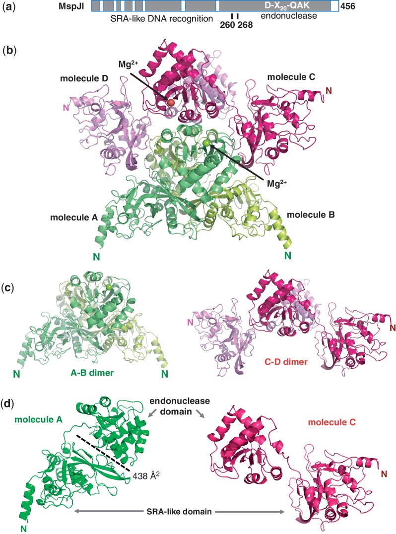
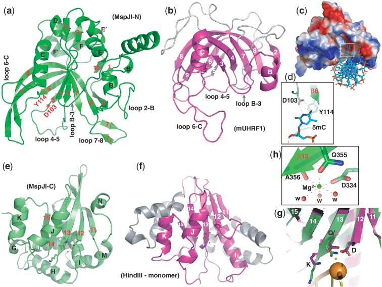
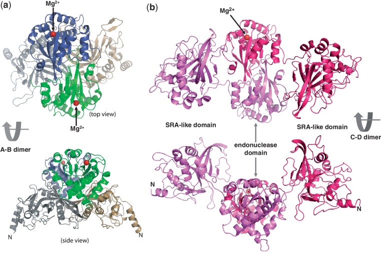
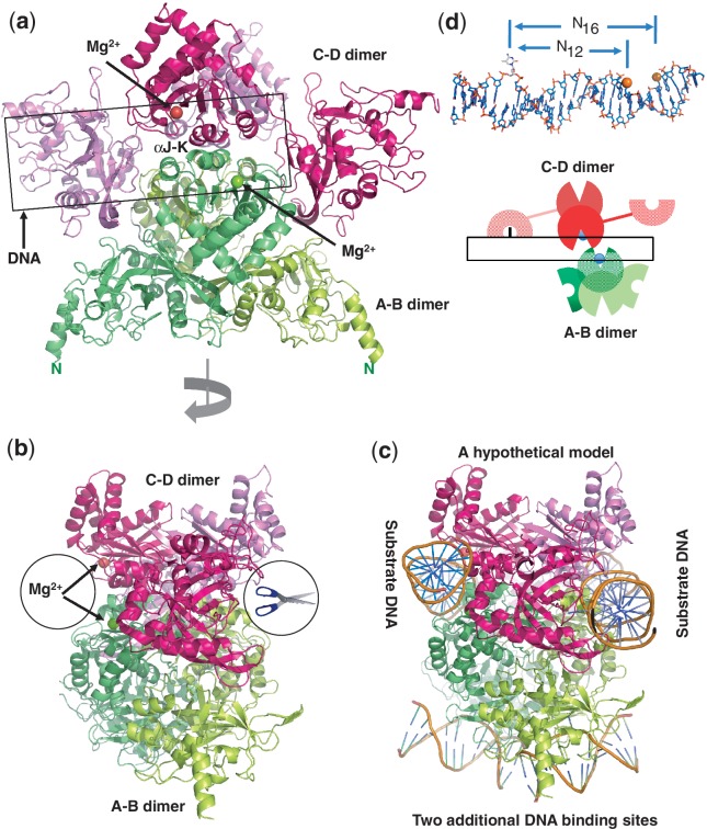
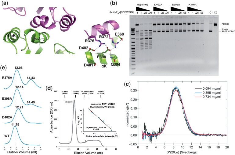
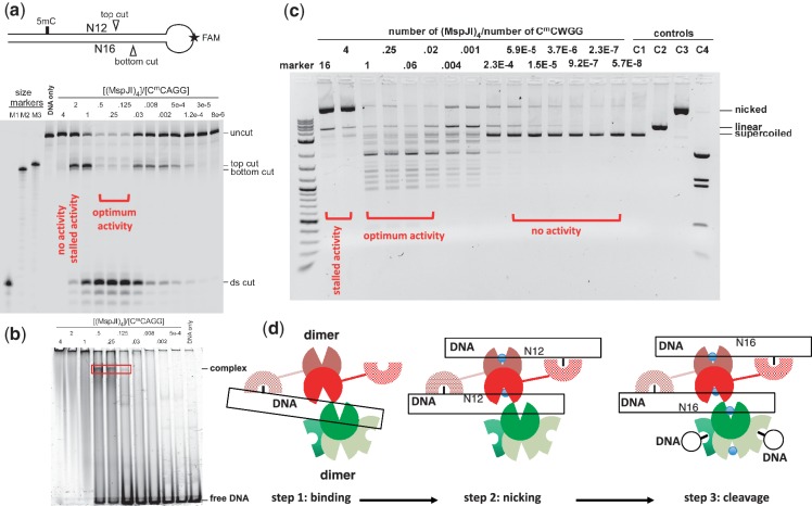
The MspJI modification-dependent restriction endonuclease recognizes 5-methylcytosine or 5-hydroxymethylcytosine in the context of CNN(G/A) and cleaves both strands at fixed distances (N(12)/N(16)) away from the modified cytosine at the 3'-side. We determined the crystal structure of MspJI of Mycobacterium sp. JLS at 2.05-Å resolution. Each protein monomer harbors two domains: an N-terminal DNA-binding domain and a C-terminal endonuclease. The N-terminal domain is structurally similar to that of the eukaryotic SET and RING-associated domain, which is known to bind to a hemi-methylated CpG dinucleotide. Four protein monomers are found in the crystallographic asymmetric unit. Analytical gel-filtration and ultracentrifugation measurements confirm that the protein exists as a tetramer in solution. Two monomers form a back-to-back dimer mediated by their C-terminal endonuclease domains. Two back-to-back dimers interact to generate a tetramer with two double-stranded DNA cleavage modules. Each cleavage module contains two active sites facing each other, enabling double-strand DNA cuts. Biochemical, mutagenesis and structural characterization suggest three different monomers of the tetramer may be involved respectively in binding the modified cytosine, making the first proximal N(12) cleavage in the same strand and then the second distal N(16) cleavage in the opposite strand. Both cleavage events require binding of at least a second recognition site either in cis or in trans.


Figures

Figure 1.
Figure 1.
Structure of MspJI. (a) Schematic representation of MspJI with two domains connected by a linker. A conserved core region is shown in dark grey and the insertions are shown as open boxes (Supplementary Figure S1). (b) Four MspJI monomers, A, B, C and D, form a tetramer. Label ‘N’ indicates amino terminus of each molecule. (c) Two kinds of dimers in closed (A–B dimer) or open (C–D dimer) conformations. (d) Two kinds of monomers with Molecules A or B adopting a closed conformation (in green) and Molecules C or D adopting an open conformation (in red).
Figure 2.
Figure 2.
The SRA-like hemi-methylated 5mC recognition domains. (a) Ribbon model of the N-terminal DNA-binding domain of MspJI. In comparison to the SRA domain of mouse UHRF1 (panel b), additional helices of MspJI are positioned on the outer surface of the crescent. In addition, loop 2–B (between strand β2 and helix αB) and loops 7–8 (between strands β7 and β8) in MspJI vary in sequence and length among family members (Supplementary Figure S1a), indicating their potential function in defining the specificity of the recognition of the DNA sequence for the nucleotides flanking of the modified cytosine. (b) The SRA domain of mouse UHRF1 (PDB 3FDE). In mammalian SRA, the corresponding loop between strand β6 and helix αC (6-C) contains residues important for CpG recognition and the loop between helix αB and strand β3 (B-3) for flipping the 5mC out of the DNA helix (22). (c) A surface model of the N-terminal SRA-like DNA recognition domain docked with a DNA containing a flipped 5mC (taken from PDB 3FDE). The surface charge is displayed as blue for positive, red for negative and white for neutral. (d) The flipped 5mC nucleotide can be docked into the binding pocket by interactions (via hydrogen bonds and planar stacking contacts) with Asp103 and Tyr114—two conserved residues among the MspJI family and known SRA domains. Asp103 is part of the loop between strand β4 and β5 (loops 4–5) and the last residue prior to strand β5. Tyr114 is part of the strand β6, which is anti-parallel to strand β5 and sits right next to Asp103. (e) Ribbon model of the C-terminal endonuclease domain of MspJI, which contains five β-strands (β11–β15) and eight helices (αG to αN). Helices αJ, αK, αN are located on one side of the β-sheet and αG, αH, αI, αL, αM on the other side, respectively. (f) The HindIII monomer (taken from the dimer-DNA complex structure; PDB 2E52). (g) Superimposition of the C-terminal endonuclease domain of MspJI (in green) and the HindIII–DNA complex (in magenta) near the scissile phosphate group (shown as an orange ball) (taken from PDB 2E52). Three catalytic residues (Asp334, Gln355 and Lys357 in MspJI and Asp93, Asp108 and Lys110 in HindIII) are shown in a stick model in the carboxyl ends of strands β12 and β13. (h) The octahedral coordination of one Mg2+ observed in the active site.
Figure 3.
Figure 3.
Two distinct back-to-back dimers of MspJI. (a) Molecules A and B form a closed back-to-back dimer with two active sites (indicated by the Mg2+ sites) located in opposite ends. Molecule A is colored in grey (the N-terminal SRA-like domain) and green (the C-terminal endonuclease domain), while Molecule B is in light orange and dark blue, respectively. (b) Molecules C and D form an open back-to-back dimer with no direct interactions between the two N-terminal DNA-binding domains.
Figure 4.
Figure 4.
A unique tetramer of MspJI. (a) Two back-to-back dimers form a tetramer with four DNA-binding domains and two face-to-face ds ‘scissors’ that cleave hexanucleotides producing four base pair staggers (N12/N16). (b) A 90° view from that of panel a. (c) A hypothetical model of MspJI with two DNA molecules bound in the active sites of the ‘scissors’ (panel b). These two DNA molecules could be connected through DNA looping. Two additional 5mC-containing DNAs could be bound through the N-terminal DNA-binding domains of A–B dimer (bottom). (d) A cartoon illustration of the proposed MspJI tetramer–DNA complex mediated by reading of 5mC by one monomer (Molecule C), cutting the proximal N12 site in the top strand by the second monomer (Molecule D) and the distal N16 cut in the bottom strand by the third monomer (Molecule A). Top panel shows a DNA molecule with a flipped 5mC and the proximal N12 (top strand) and distal N16 (bottom strand) cleavage sites.
Figure 5.
Figure 5.
MspJI exists as a tetramer in solution. (a) The major tetramer interface is mediated by helices αJ and αK (left panel), including a network of charge–charge interactions (right panel). (b) Activity profiles of the mutants D402A, E398A and R376A showing the cleavage stalled in the nicked state, compared with the MspJI wild-type. The 4-fold titrations of MspJI and mutants were incubated with 200 ng of pBR322 at 37°C for 2 h. (c) Analytical ultracentrifugation of MspJI at three different concentrations. Scans were taken every 4.5 min and were used to calculate the normalized sedimentation coefficient distribution, g(s*). The tetramer has a sedimentation coefficient corrected to standard conditions, S(20,w), of 8.64 S. (d) Elution profile of MspJI on a Superdex 200 (10/300 GL) (GE Healthcare). The column buffer was 20 mM Tris, pH 8.0, 1 mM EDTA, 10% glycerol (v/v), 1 mM DTT and 150 mM NaCl, and ∼1.7 mg of MspJI was loaded onto the column. The inset shows the standardization of the size exclusion column using a protein marker kit (Biorad) at the time MspJI was profiled using the same buffer. (e) Elution profiles of MspJI mutants (D402A, E398A and R376A) and wt on a Superdex 200 (10/300 GL). The column buffer was the same as in panel d and ∼100 µg of protein was loaded onto the column in four consecutive runs.
Figure 6.
Figure 6.
MspJI cleaves via a top-strand nicked intermediate. (a) A stem-loop structured oligonucleotide substrate containing one 5mC site was designed with an internal fluorescent FAM label in the loop. Size markers were synthesized according to predicted cleavage sites: M1, product from a ds cleavage; M2, product from the bottom-strand cleavage; M3, product from the top-strand cleavage. A 2-fold titration of MspJI digestion started from the tetramer to DNA ratio of 4 to 0.125 and followed by 4-fold dilution. (b) DNA-binding assays were performed by incubating 0.5 µM FAM labeled stem-loop oligonucleotides with varying amount of MspJI tetramer at 37°C for 1 h. (c) MspJI titration on pBR322 (dcm+) containing six C5mCWGG sites. The molar ratio of MspJI tetramer to its substrate sites is shown on the top of the lanes. Control lanes include: C1, pBR322 only; C2, pBR322 digested with EcoRI, which produces linearized pBR322; C3, pBR322 digested with nicking endonuclease Nt.BspQI, which produces a nicked pBR322; C4, pBR322 digested with BstNI, which produces a complete digestion pattern at C5mCWGG. (d) A proposed three-step mechanism of the MspJI enzymatic reaction. Step 1: one SRA-like domain binds specifically to the modified cytosine. With a tetramer-to-DNA ratio of 4:1, no enzymatic activity was observed. Step 2: the other SRA-like domain of the same back-to-back dimer binds another target site, resulting in a top-strand nicked intermediate. With a tetramer-to-DNA ratio of 2:1, the reaction stalled after the first N12 cut. Step 3: with a tetramer-to-DNA ratio of 1:4, the highest level of ds cleavage was observed.

References

    1. Bestor T, Laudano A, Mattaliano R, Ingram V. Cloning and sequencing of a cDNA encoding DNA methyltransferase of mouse cells. The carboxyl-terminal domain of the mammalian enzymes is related to bacterial restriction methyltransferases. J. Mol. Biol. 1988;203:971–983. - PubMed
    1. Okano M, Xie S, Li E. Cloning and characterization of a family of novel mammalian DNA (cytosine-5) methyltransferases. Nat. Genet. 1998;19:219–220. - PubMed
    1. Wu H, Zhang Y. Mechanisms and functions of Tet protein-mediated 5-methylcytosine oxidation. Genes Dev. 2011;25:2436–2452. - PMC - PubMed
    1. Bhutani N, Burns DM, Blau HM. DNA demethylation dynamics. Cell. 2011;146:866–872. - PMC - PubMed
    1. Williams K, Christensen J, Helin K. DNA methylation: TET proteins-guardians of CpG islands? EMBO Rep. 2011;13:28–35. - PMC - PubMed

Publication types

Substances