

doi: 10.3892/ol.2011.397.
Epub 2011 Aug 29.
Primary retroperitoneal malignant melanoma: A case report
Affiliations
- PMID: 22848275
- PMCID: PMC3406492
- DOI: 10.3892/ol.2011.397
Item in Clipboard
Primary retroperitoneal malignant melanoma: A case report
Oncol Lett.
2011 Nov.
Abstract
Primary malignant melanoma occurring at an extra cutaneous site is rare. A case of primary malignant melanoma located in the retroperitoneum of an 18-year-old female is presented in this study. Histopathological examination of the tissue biopsies at laparotomy with immunohistochemical stains confirmed a diagnosis of malignant melanoma. Further extensive clinical and radiological investigations proved the retroperitoneum to be the primary site.
Figures
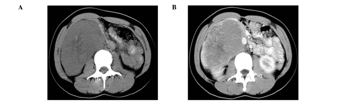
(A) Precontrast-enhanced and (B) postcontrast-enhanced CT images show a retroperitoneal mass with heterogeneous enhancement and tortuous blood vessels surrounding it.

(A) H&E, magnification, ×100. (B) H&E, magnification, ×400. A photomicrograph of the operated specimen shows epithelioid neoplastic cells with abundant eosinophilic cytoplasm, round to ovoid nuclei and prominent nucleoli, arranged in a uniform nested to fascicular pattern of growth, separated by vascularized fibrocollagenous septa (white arrow).

Immunohistochemistry of the specimen shows cells staining positive for (A) HMB-45 (magnification, ×400), (B) S-100 (magnification, ×400) and (C) melanin A (magnification, ×400).
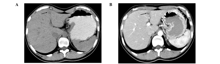
(A) Precontrast-enhanced and (B) postcontrast-enhanced CT images, taken 9 months after the patient was discharged, demonstrated a metastatic focus (black arrow).
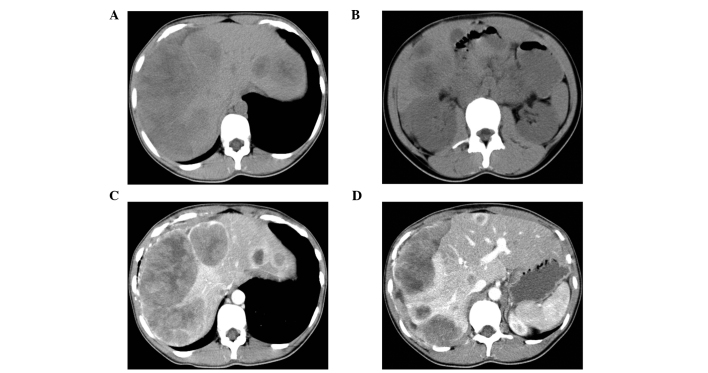
(A and B) Precontrast-enhanced CT images of different sections and (C and D) corresponding postcontrast-enhanced CT images, taken in February 2008, show the metastasis to have progressed, compared to previous scans.
Similar articles
-
Primary Retroperitoneal Melanoma Presented in a Rare Extracutaneous Site for Malignant Melanoma.Rare Tumors. 2016 Oct 5;8(3):6308. doi: 10.4081/rt.2016.6308. eCollection 2016 Sep 5. Rare Tumors. 2016. PMID: 27746882 Free PMC article.
-
A Rare Presentation of Melanoma as a Retroperitoneal Mass Seen on FDG PET.Clin Nucl Med. 2020 Apr;45(4):324-325. doi: 10.1097/RLU.0000000000002920. Clin Nucl Med. 2020. PMID: 31977477
-
Primary malignant melanoma of the small intestine: a case report.Acta Chir Belg. 2009 May-Jun;109(3):405-7. doi: 10.1080/00015458.2009.11680448. Acta Chir Belg. 2009. PMID: 19943602
-
Primary malignant melanoma of the ovary: case report and review of the literature.Turk Patoloji Derg. 2011 May;27(2):169-72. doi: 10.5146/tjpath.2011.01069. Turk Patoloji Derg. 2011. PMID: 21630207 Review.
-
Role of In Vivo Reflectance Confocal Microscopy in the Analysis of Melanocytic Lesions.Acta Dermatovenerol Croat. 2018 Apr;26(1):64-67. Acta Dermatovenerol Croat. 2018. PMID: 29782304 Review.
Cited by
-
Primary Retroperitoneal Malignant Melanoma with Involvement of Iliac Artery and Vein.Case Rep Med. 2021 Sep 4;2021:3526071. doi: 10.1155/2021/3526071. eCollection 2021. Case Rep Med. 2021. PMID: 34527055 Free PMC article.
-
Primary Retroperitoneal Melanoma Presented in a Rare Extracutaneous Site for Malignant Melanoma.Rare Tumors. 2016 Oct 5;8(3):6308. doi: 10.4081/rt.2016.6308. eCollection 2016 Sep 5. Rare Tumors. 2016. PMID: 27746882 Free PMC article.
References
-
- Rager EL, Bridgeford EP, Ollila DW. Cutaneous melanoma: Update on prevention, screening, diagnosis, and treatment. Am Fam Physician. 2005;72:269–276. - PubMed
-
- Chang AE, Karnell LH, Menck HR. The National Cancer Data Base report on cutaneous and noncutaneous melanoma: a summary of 84,836 cases from the past decade. The American College of Surgeons Commission on Cancer and the American Cancer Society. Cancer. 1998;83:1664–1678. - PubMed
-
- Capizzi P, Donohue J. Metastatic melanoma of the gastrointestinal tract: a review of the literature. Compr Ther. 1994;20:20–23. - PubMed
-
- Poos HPAM, Kruijff S, Bastiaannet E, Van Ginker RJ, Hoekstra HJ. Therapeutic groin dissection for melanoma: Risk factors for short term morbidity. Eur J Surg Oncol. 2009;35:877–883. - PubMed
LinkOut - more resources
Full Text Sources