. 2012;7(7):e40401.
doi: 10.1371/journal.pone.0040401. Epub 2012 Jul 27.

Serratia odorifera a midgut inhabitant of Aedes aegypti mosquito enhances its susceptibility to dengue-2 virus

Affiliations

Serratia odorifera a midgut inhabitant of Aedes aegypti mosquito enhances its susceptibility to dengue-2 virus

Anjali Apte-Deshpande et al. PLoS One. 2012.

Abstract

Mosquito midgut plays a crucial role in its vector susceptibility and pathogen interaction. Identification of the sustainable microflora of the midgut environment can therefore help in evaluating its contribution in mosquito-pathogen interaction and in turn vector competence. To understand the bacterial diversity in the midgut of Aedes aegypti mosquitoes, we conducted a screening study of the gut microbes of these mosquitoes which were either collected from fields or reared in the laboratory "culture-dependent" approach. This work demonstrated that the microbial flora of larvae and adult Ae. aegypti midgut is complex and is dominated by gram negative proteobacteria. Serratia odorifera was found to be stably associated in the midguts of field collected and laboratory reared larvae and adult females. The potential influence of this sustainable gut microbe on DENV-2 susceptibility of this vector was evaluated by co-feeding S. odorifera with DENV-2 to adult Ae. aegypti females (free of gut flora). The observations revealed that the viral susceptibility of these Aedes females enhanced significantly as compared to solely dengue-2 fed and another gut inhabitant, Microbacterium oxydans co-fed females. Based on the results of this study we proposed that the enhancement in the DENV-2 susceptibility of Ae. aegypti females was due to blocking of prohibitin molecule present on the midgut surface of these females by the polypeptide of gut inhabitant S. odorifera.


Conflict of interest statement

Competing Interests: The authors have declared that no competing interests exist.

Figures

Figure 1
Figure 1. Serratia odorifera and Ae. aegypti interaction.
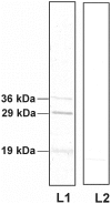
a. Significance of S. odorifera’s presence in the blood meal on the DENV-2 susceptibility of Ae. aegypti: Adult females were fed with Blood + DENV-2, Blood + DENV-2+ M. oxydans (Blood + DENV-2+ S. odorifera via blood meal. DENV-2 dissemination was detected in the head squashes on 14 post infection days by IFA. The presence of S. odorifera in the blood meal significantly enhanced the dengue virus susceptibility (Mann-Whitney U test; P<0.05) compared to M. oxydans (Mann-Whitney U test; P>0.05). Post feeding virus titers in the blood meal were determined by plaque assay (1.8× 10⁵ PFU/mL of blood). b. Overlay assay with S. odorifera and M. oxydans cell lysates: The bacterial cell lysates of S. odorifera (So1 and So2) and M. oxydans (Mo1 and Mo2) were separated by SDS-PAGE and transferred to a nitrocellulose membrane. The membrane was overlaid with BBMF of Ae. aegypti. The putative binding proteins were detected by mouse anti-BBMF antibody and HRP labeled-secondary antibody. c. Expression of P40 in cell lysates and cell supernatants under different temperature conditions: S. odorifera cell lysate (S1 and S3 30°C, S2 41°C), M. oxydans cell lysate (M1 and M3 30°C, M2 41°C), culture filtrate of S. odorifeara (S4 and S6 30°C, S5 41°C) and culture filtrate of M. oxydans (M4 and M6 30°C, M5 41°C) were separated by SDS-PAGE and transferred to Hybond-C membranes. The membranes were incubated with the anti P40 mouse IgG (lanes S1, S2, S4, S5, M1, M2, M4 and M5) and with PBS pH 7.4 (lanes S3, S6, M3 and M6). Presence of P40 was detected by incubating the membranes with the secondary antibody (peroxidase-conjugated goat anti mouse IgG). Reaction was developed using H2O2 and DABT.d. Protein-protein interaction between BBMF and S. odorifera cell lysate: The membrane proteins of Ae. aegypti midgut (Lanes L1, L2 ) were separated by SDS–PAGE and transferred to Hybond-C membranes. The membranes were incubated with S. odorifera cell lysate (L1) and PBS pH 7.4 (L2) at 37°C. The putative P40 binding proteins were visible after incubation with anti P40 mouse antibody and the secondary antibody (peroxidase-conjugated goat anti mouse IgG). The reaction was developed using H2O2 and DABT. The molecular weights of DENV-2 binding proteins are shown on the left side of the blot.
Figure 2
Figure 2. P40 localization in the Ae. aegypti gut.
The midgut sections (10 µm) of Ae. aegypti fourth instar larvae (a) adult female (b) and slit opened gut of adult females (c) were incubated with S. odorifera cell lysate and control midgut sections were incubated with PBS (pH 7.4). P40 interaction with the midgut epithelium was detected using mouse anti-P40 antibody and with a Cy3 conjugated rabbit anti mouse IgG secondary antibody. The signal was detected using a Zeiss microscope equipped with the Axiovesion detection system.
Figure 3
Figure 3. Virus overlay protein binding assay (VOPBA).
Serratia odorifera cell lysate (lanes L1 and L2) were subjected to SDS-PAGE and transferred to Hybond-C membranes. Lane L1 was incubated with 5×10⁵ plaque-forming units of DENV-2, and lane L2 with PBS (pH 7.4) at 37°C. The putative DENV-2 binding proteins were visible after incubation with a rabbit polyclonal antibody to DENV-2 and a secondary antibody (peroxidase-conjugated goat anti-rabbit IgG). The reaction was developed using H2O2 and DABT. The molecular weights of dengue-2 binding proteins are shown on the left side of the blot.

References

    1. Dillon RJ, Vennard CT, Buckling A, Charnley AK (2005) Diversity of locust gut bacteria protects against pathogen invasion. Ecol Lett 8: 1291–1298.
    1. Dong Y, Manfredini F, Dimopoulos G (2009) Implication of the Mosquito Midgut Microbiota in the defence against Malaria Parasites. PLoS Pathog 5: e1000423 doi:10.1371/journal.ppat.1000423. - DOI - PMC - PubMed
    1. Jadin J, Vincke IH, Dunjic A, Delville JP, Wery M, et al. (1966) Role of Pseudomonas in the sporogenesis of the hematozoon of malaria in the mosquito. Bull Soc Pathol Exot Filiales 59: 514–525. - PubMed
    1. Seitz HM, Maier WA, Rottok M, Becker-Feldmann H (1987) Concomitant infections of Anopheles stephensi with Plasmodium berghei and Serratia marcescens: additive detrimental effects. Zentralbl Bakteriol Hyg 266: 155–166. - PubMed
    1. Pumpuni CB, DeMaio J, Kent M, Davis JR, Beier JC (1996) Bacterial population dynamics in three anopheline species: the impact on Plasmodium sporogonic development. Am J Trop Med Hyg 54: 214–218. - PubMed

Publication types