. 2012;7(7):e40795.
doi: 10.1371/journal.pone.0040795. Epub 2012 Jul 27.

Hsp90 is cleaved by reactive oxygen species at a highly conserved N-terminal amino acid motif

Affiliations

Hsp90 is cleaved by reactive oxygen species at a highly conserved N-terminal amino acid motif

Raphaël Beck et al. PLoS One. 2012.

Abstract

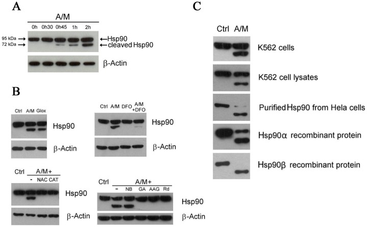
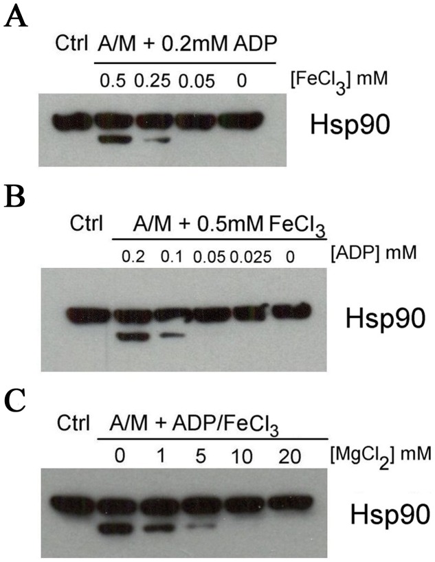
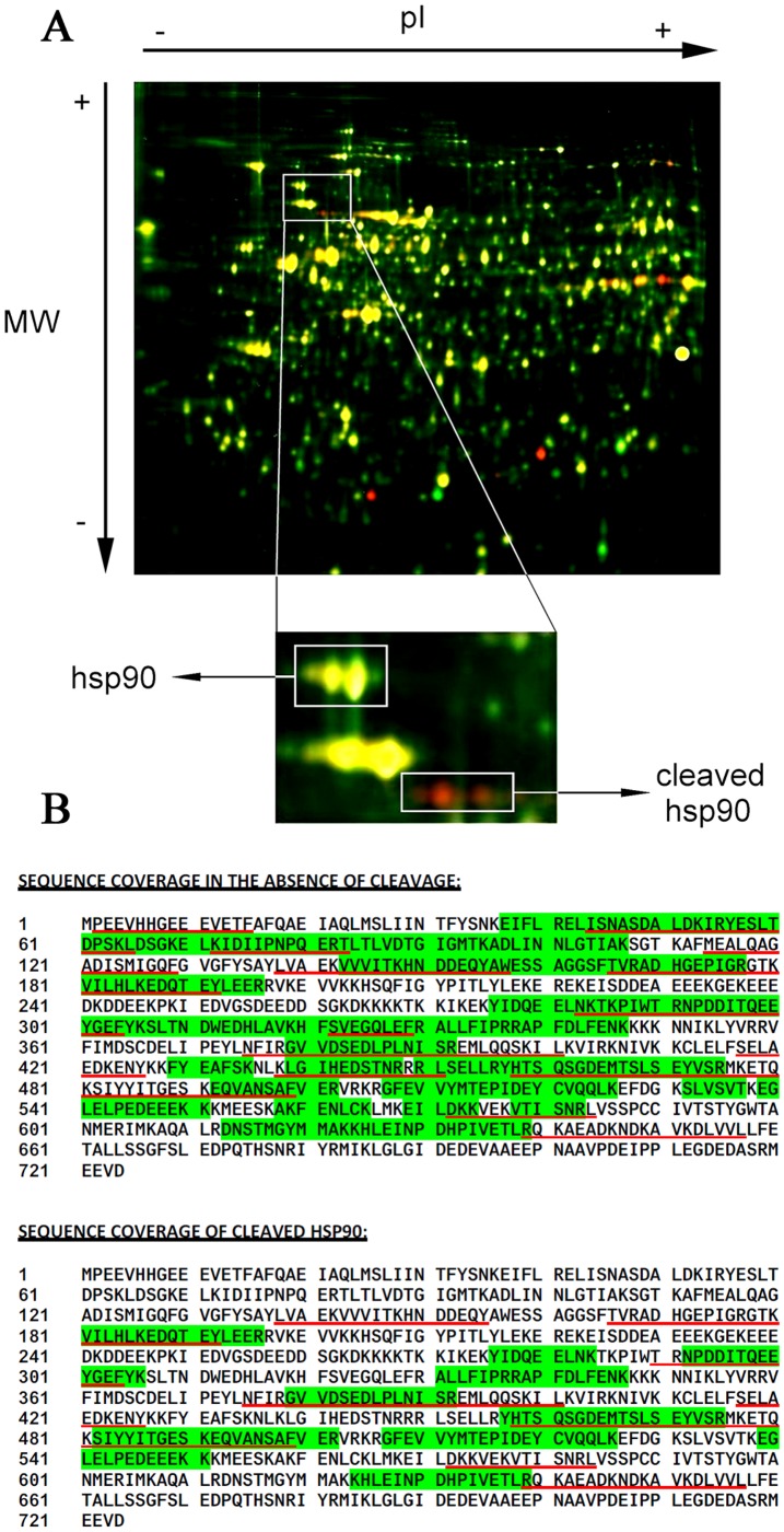
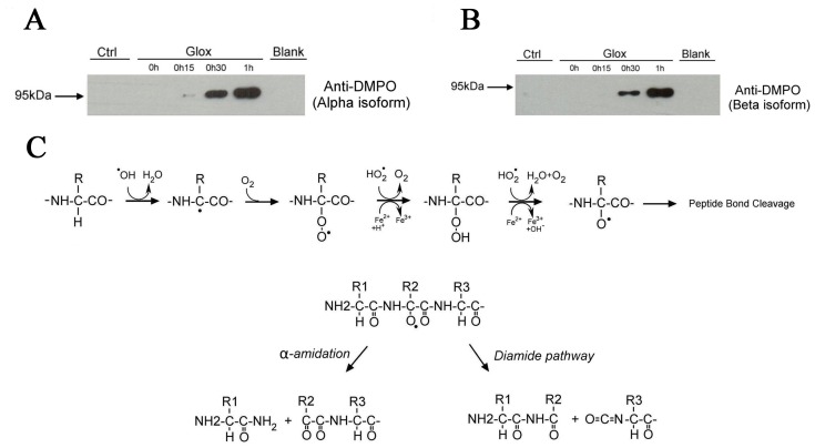
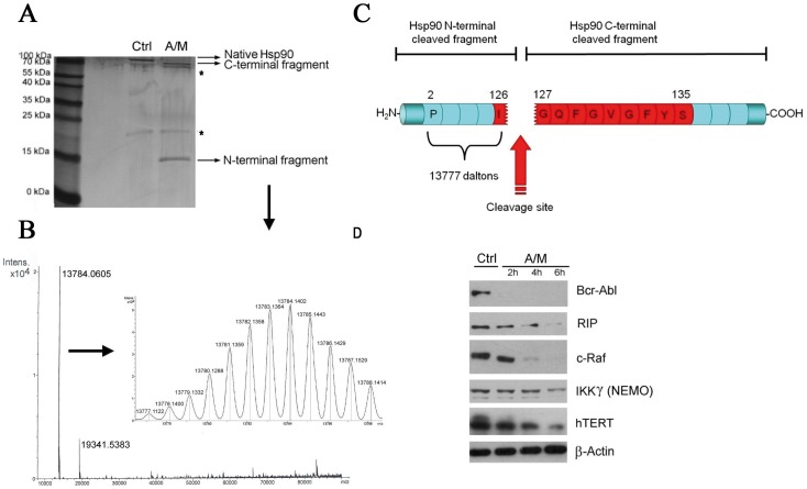
Hsp90 is an essential chaperone that is necessary for the folding, stability and activity of numerous proteins. In this study, we demonstrate that free radicals formed during oxidative stress conditions can cleave Hsp90. This cleavage occurs through a Fenton reaction which requires the presence of redox-active iron. As a result of the cleavage, we observed a disruption of the chaperoning function of Hsp90 and the degradation of its client proteins, for example, Bcr-Abl, RIP, c-Raf, NEMO and hTert. Formation of Hsp90 protein radicals on exposure to oxidative stress was confirmed by immuno-spin trapping. Using a proteomic analysis, we determined that the cleavage occurs in a conserved motif of the N-terminal nucleotide binding site, between Ile-126 and Gly-127 in Hsp90β, and between Ile-131 and Gly-132 in Hsp90α. Given the importance of Hsp90 in diverse biological functions, these findings shed new light on how oxidative stress can affect cellular homeostasis.


Conflict of interest statement

Competing Interests: The authors have declared that no competing interests exist.

Figures

Figure 1
Figure 1. Oxidative stress leads to Hsp90 cleavage and client protein degradation.
(A) Time course of Hsp90 cleavage in K562 cells upon treatment with A/M (2 mM/10 µM). Hsp90 was detected with an anti-C terminus antibody from Santa Cruz Biotechnology (Hsp90 α/β, clone F-8). (B) Hsp90 cleavage in cells incubated for 2 hours with either A/M (2 mM/10 µM) or Glox (30 mM glucose/0.25 U/ml glucose oxidase), another H2O2-generating system (top left). Suppression of Hsp90 cleavage in cells preincubated for 1 h with 3 mM N-acetylcysteine (NAC) or 100 U/ml of catalase (CAT) and then exposed to A/M for 2 h (bottom left). Iron chelation by preincubating cells with deferoxamine (DFO) for 18 h decreased the cleavage induced by 2 h treatment with A/M (top right). Preincubation of cells for 1 h with 10 µM of N-term Hsp90 inhibitors, like geldanamycin (GA), 17-AAG (AAG) or radicicol (Rd), protected Hsp90 from the cleavage induced by 2 h treatment with A/M. Novobiocin (NB) at 1 mM did not protect (bottom right). Hsp90 was detected with the same antibody as in (A). (C) Cellular Hsp90 cleavage was reproduced in K562 cell lysates, in purified Hsp90 from HeLa cells, and in Hsp90 α and β recombinant proteins. K562 cells were incubated for two hours in the absence (Ctrl) or in the presence of A/M (2 mM/10 µM). K562 cell lysates (100 µg) were incubated for 1 h in the absence (Ctrl) or in the presence of A/M supplemented with ADP (0.2 mM) and FeCl3 (0.5 mM). For the experiments with purified and recombinant proteins, we used 2 µg of Hsp90, incubation lasted 30 min and A/M (2 mM/10 µM) was supplemented with 0.2 mM ADP and 0.5 mM FeCl3. Note that Hsp90 was detected with the same antibody as in (A) with the exception of recombinant Hsp90α that was detected with an anti-penta-His antibody.
Figure 2
Figure 2. Hsp90 cleavage by A/M requires the presence of ionic iron and ADP.
K562 cell lysates (100 µg) were incubated for 1 h in the absence (Ctrl) or in the presence of A/M (2 mM/10 µM) supplemented with ADP (0.2 mM) and FeCl3 (0.5 mM). Different concentrations of ADP (A), FeCl3 (B) and MgCl2 (C) were tested, as indicated. Hsp90 was detected with an anti-C terminus antibody from Santa Cruz Biotechnology (Hsp90 α/β, clone F-8).
Figure 3
Figure 3. 2D-DIGE analysis of protein extracts from K562 cells exposed to oxidative stress.
(A) K562 cells incubated for 2 h in the absence (green) or the presence (red) of A/M (2 mM/10 µM). Spots corresponding to Hsp90 and its cleaved fragments were excised and analyzed by mass spectrometry. (B) Peptides found after mass spectrometry analysis of the excised 2D-DIGE spots of cleaved and non-cleaved Hsp90 protein by either tryptic (highlighted in green) or chymotryptic (underlined in red) peptides. Alignment was performed against the Hsp90β protein sequence.
Figure 4
Figure 4. Formation of Hsp90 protein radicals following oxidative stress.
(A) Immuno-spin trapping blots showing the formation of nitrone adducts in Hsp90α (200 µg) incubated with DMPO (1 mM) and exposed to oxidative stress generated by Glox (60 mM glucose/1 U/ml glucose oxidase) supplemented with ADP (0.2 mM) and FeCl3 (0.5 mM). Blank and Ctrl mean, respectively, that DMPO and Glox were omitted. (B) Same as (A) but with Hsp90β. (C) Proposed mechanism leading to oxidative polypeptide cleavage according to Stadman et al , .
Figure 5
Figure 5. Identification of the site of cleavage within Hsp90.
(A) Silver stained SDS-PAGE gel showing both C-term and N-term protein fragments of Hsp90β after 30 min incubation with A/M (2 mM/10 µM) supplemented with ADP (0.2 mM) and FeCl3 (0.5 mM). Asterisks show the presence of contaminants. (B) Mass spectrometry analysis of cleaved Hsp90β. Deconvoluted spectra of the small N-term fragment gave a precise monoisotopic mass of 13777.1122 daltons. (C) The picture illustrates the location of the cleavage site occurring in Hsp90β. The IGQFGVGFYS motif corresponding to a conserved amino acid sequence in several Hsp90 proteins is highlighted in red. (D) Degradation of various Hsp90 client proteins in K562 cells treated with A/M (2 mM/10 µM).

References

    1. Borkovich KA, Farrelly FW, Finkelstein DB, Taulien J, Lindquist S. hsp82 is an essential protein that is required in higher concentrations for growth of cells at higher temperatures. Mol Cell Biol. 1989;9:3919–3930. - PMC - PubMed
    1. Pearl LH, Prodromou C. Structure and mechanism of the Hsp90 molecular chaperone machinery. Annu Rev Biochem. 2006;75:271–294. - PubMed
    1. McLaughlin SH, Ventouras LA, Lobbezoo B, Jackson SE. Independent ATPase activity of Hsp90 subunits creates a flexible assembly platform. J Mol Biol. 2004;344:813–826. - PubMed
    1. Neckers L. Hsp90 inhibitors as novel cancer chemotherapeutic agents. Trends Mol Med. 2002;8:S55–61. - PubMed
    1. Schneider C, Sepp-Lorenzino L, Nimmesgern E, Ouerfelli O, Danishefsky S, et al. Pharmacologic shifting of a balance between protein refolding and degradation mediated by Hsp90. Proc Natl Acad Sci U S A. 1996;93:14536–14541. - PMC - PubMed

Publication types